Figure 5. Ultrastructure of the outer retina in EE and ST mice. Electron micrographs of the outer retina of EE (A, C) and ST (B) rd10 mice. Asterisks in the enriched environment (EE) sample label nuclei of residual cells in the outer retina, adjacent
to a well-organized outer limiting membrane (OLM). These nuclei have different densities and presumably belong to surviving
cones and misplaced bipolar cells. The outer plexiform layer (OPL) is still present in A and C, while in the ST sample (D), the cell bodies of the inner nuclear layer (INL) cells are in close proximity to the RPE.
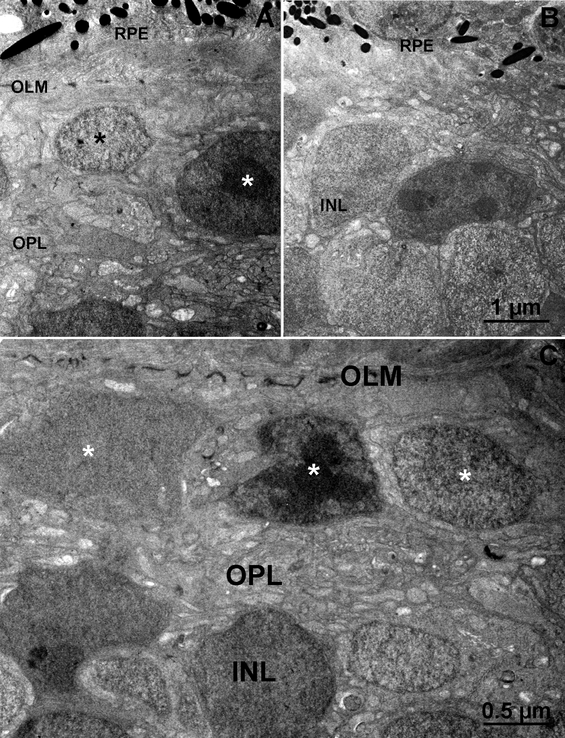
 Figure 5 of
Barone, Mol Vis 2014; 20:1545-1556.
Figure 5 of
Barone, Mol Vis 2014; 20:1545-1556.