Figure 4. The outer retina in in EE and ST mice. Semithin (2 µm thick) plastic sections from the retinas of rd10EE (A) and rd10 ST (B) mice, obtained at similar locations. The retina in A is thicker and shows a layer of scattered cell bodies (arrows) abutting the RPE. In A and B, sparse granules of RPE pigment (visible as bright, elongated drops) penetrate the neural retina. INL = inner nuclear layer;
IPL = inner plexiform layer.
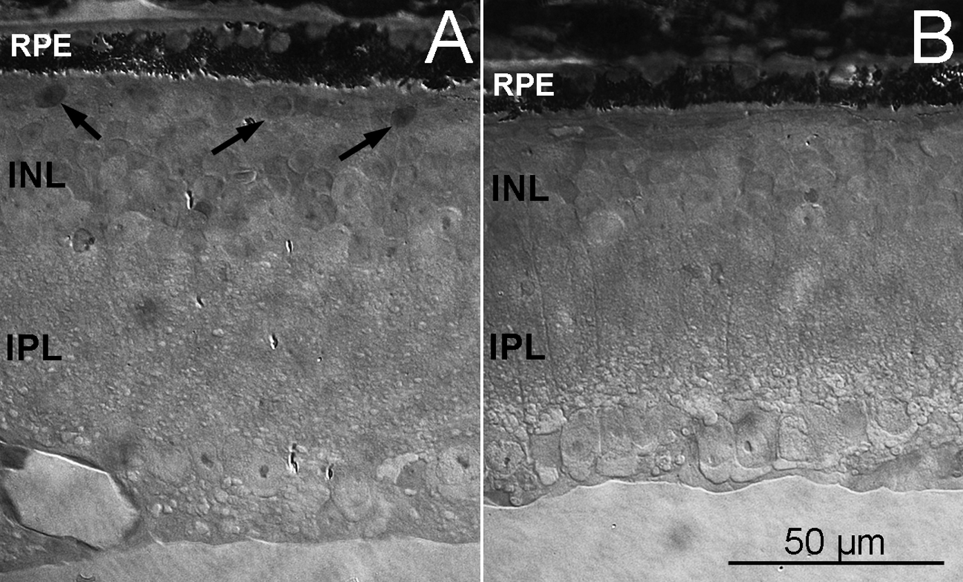
 Figure 4 of
Barone, Mol Vis 2014; 20:1545-1556.
Figure 4 of
Barone, Mol Vis 2014; 20:1545-1556.