Figure 3. Cone survival in in EE and ST mice. A and B: Retinal whole mounts from 1-year-old rd10 mice raised in EE (A) and ST conditions (B), stained with antibodies against cone-specific opsins (red signal). A large black area devoid of cones is evident in the
central part of the retina, revealing a nasal-temporal pattern of degeneration. Cone clusters (visible as red, punctate areas)
are still preserved in the periphery of both retinas. These low magnification images show retinal tears that form easily in
the standard laboratory (ST) samples during dissection (B). D, V, N, T: dorsal, ventral, nasal, temporal retinal poles. C and D: High-magnification confocal images of cones obtained from the retinal samples shown in A and B, at corresponding locations. Local density of cones is visibly higher in the enriched environment (EE) compared to the ST
sample. Typical cone remodeling is evident in both cases, but the EE cones display apical protrusions reminiscent of residual
outer segments (arrow). E: Counts from four retinas in each group show that the rd10 EE mice at 1 year have almost three times as many surviving cones
(34,000±4,000) as the ST control mice (12,700±1,800) t test, **p=0.003.
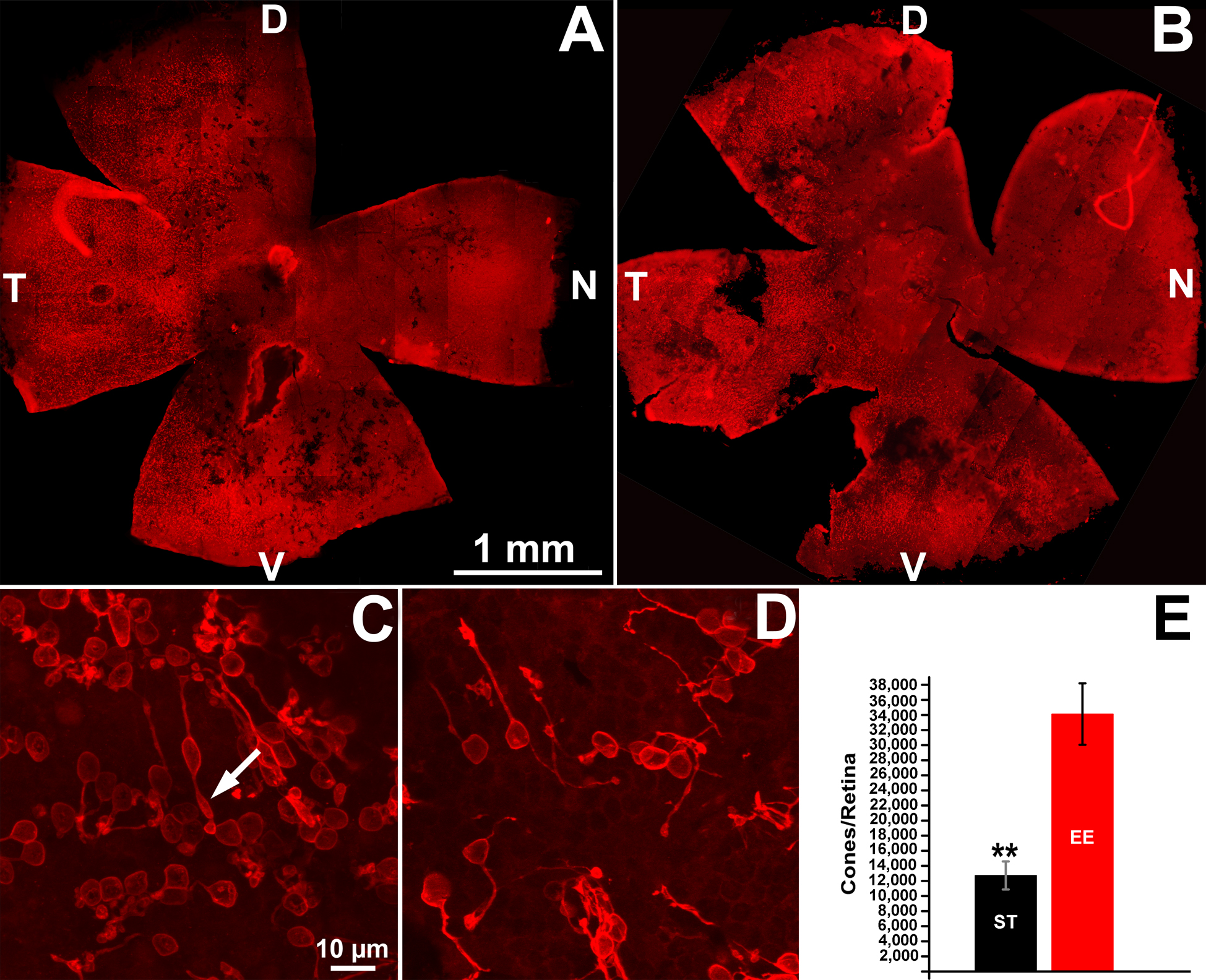
 Figure 3 of
Barone, Mol Vis 2014; 20:1545-1556.
Figure 3 of
Barone, Mol Vis 2014; 20:1545-1556.