Figure 1. Retinal degeneration in rd10 EE and ST mice. Fluorescence nuclear staining of vertical retinal sections obtained from a WT
mouse (A), an rd10 EE mouse (B), and an rd10 ST mouse (C), all aged 1 year. Extensive photoreceptor degeneration with the disappearance of the outer nuclear layer (ONL) is clear
in B and C. The sample in C also shows highly irregular outer and inner retinal margins, typical of the extremely thin and fragile retinas of rd10 ST
mice of this age. INL = inner nuclear layer; IPL = inner plexiform layer.
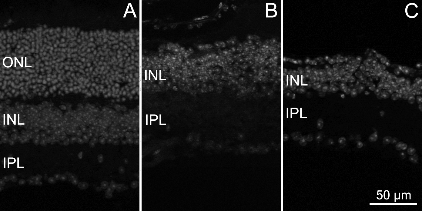
 Figure 1 of
Barone, Mol Vis 2014; 20:1545-1556.
Figure 1 of
Barone, Mol Vis 2014; 20:1545-1556.