Figure 4. Daily systemic valproic acid (VPA; P9–P21, early treatment) reduced the rate of photoreceptor loss in Pde6brd1/rd1 mice. A: Systemic VPA treatment of Pde6brd1/rd1 mice was 350 mg/kg/day (IP injection) from age P9 to P21 (early treatment). Three to four extra layers of photoreceptor cells
(nuclei) remained in VPA-treated mice compared to PBS-treated controls. Examples of retinal sections (P21) are shown from
a VPA-treated and PBS-treated littermate (control). White arrows indicate the outer nuclear layer (ONL). Regions of the ONL
just left of the white arrows are also shown below at higher magnification (sections are paraffin, hematoxylin and eosin stained).
B: Spiderogram graphs showing the thicker ONL in Pde6brd1/rd1 mice after daily systemic VPA early treatment (P9–P21) compared to PBS-treated littermates. ONL thickness is plotted at 200-µm
intervals from the optic nerve to the retinal periphery, using a virtual microscopy of full retinal histology sections (paraffin,
hematoxylin and eosin stained; n = 3/group, t test, *p<0.05, **p<0.01, relative to PBS). C: Systemic VPA treatment of wild-type mice from age P9 to P21 (early treatment) resulted in a 10%–20% loss of photoreceptor
cells. Example SD-OCT images (P21) and retinal histology sections (P21) from the same VPA-treated and PBS-treated littermates
are shown, confirming thinning of the ONL in vivo (INL, inner nuclear layer; OLM, outer limiting membrane). D: Spiderogram graphs showing the thinner ONL in wild-type mice after daily systemic VPA early treatment (P9–P21) compared
to PBS-treated littermates (n = 4/group, t test, *p<0.05, **p<0.01, relative to PBS).
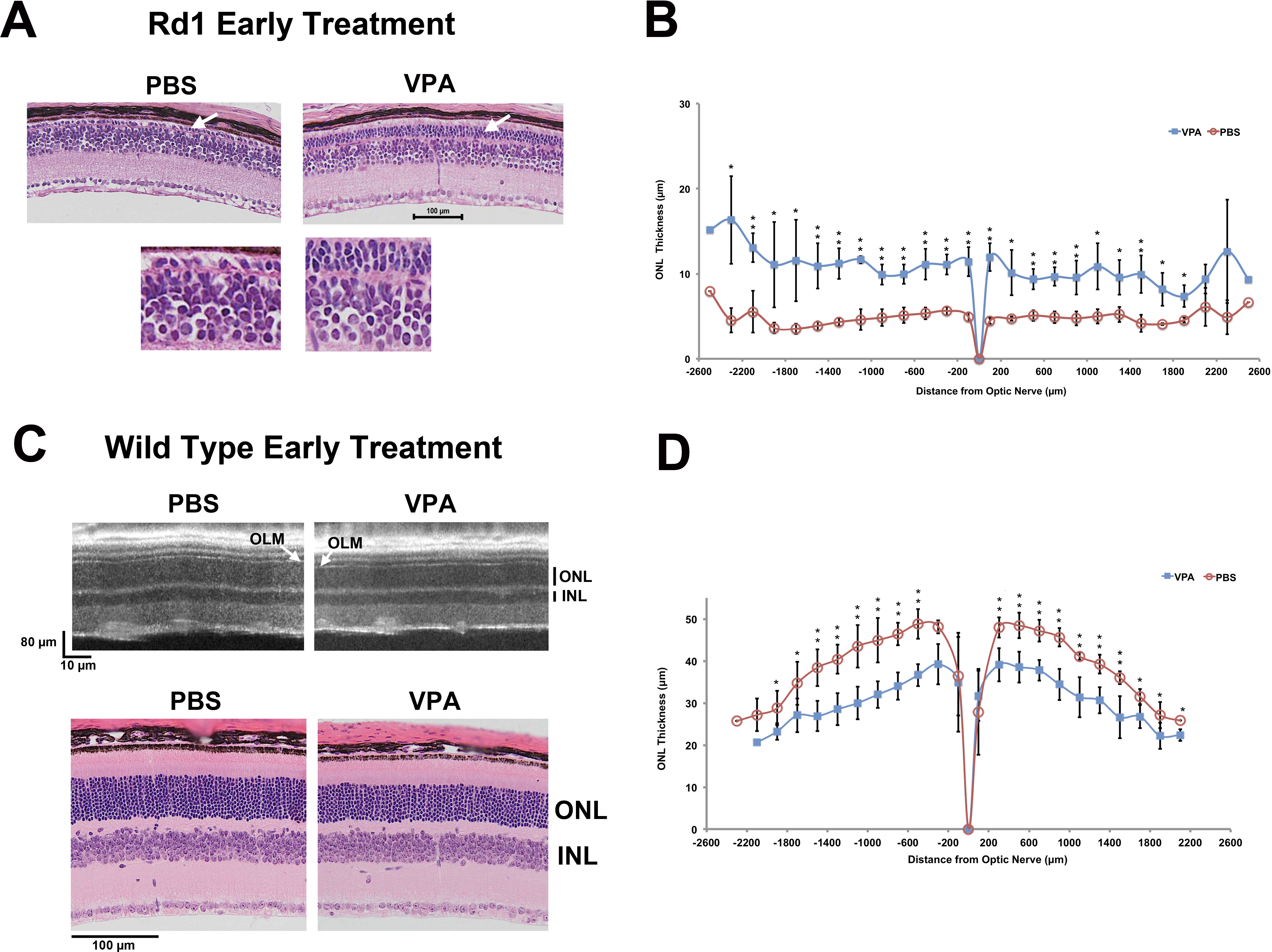
 Figure 4 of
Mitton, Mol Vis 2014; 20:1527-1544.
Figure 4 of
Mitton, Mol Vis 2014; 20:1527-1544.