Figure 4. Immunohistochemistry of the retina of rats with experimental autoimmune uveoretinitis. Representative results for expression
of vascular endothelial growth factor (VEGF; green) and glutamine synthetase (GS; red) are shown on day 28 post-immunization
treated with vehicle (A), low-dose fenofibrate (FF(20); B), and high-dose fenofibrate (FF(100); C). Increased expression of VEGF was observed in Müller cells (A and B). Rats treated with high-dose fenofibrate showed faint expression of VEGF in Müller cells and a marked decrease in ocular
infiltrative cells (C). The bar represents 50 µm.
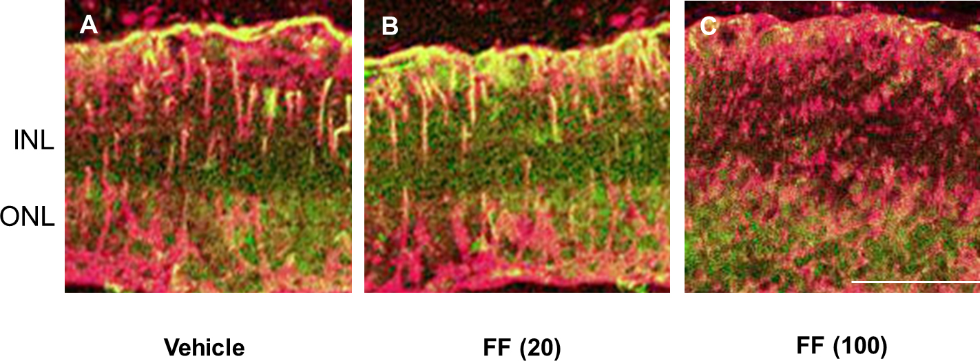
 Figure 4 of
Osada, Mol Vis 2014; 20:1518-1526.
Figure 4 of
Osada, Mol Vis 2014; 20:1518-1526.