Figure 3. Immunohistochemistry of retinal sections from rats with experimental autoimmune uveoretinitis treated with vehicle (n=4),
low-dose fenofibrate (n=4), and high-dose fenofibrate (n=4) on day 28 post-immunization. Marked expression of interleukin
(IL)-6 (green) and IL-17 (red) was observed in ocular infiltrative cells in rats treated with vehicle (A). Rats treated with a high dose of fenofibrate showed weak expression of IL-6 and IL-17, and marked decrease in ocular infiltrative
cells. INL; inner nuclear layer, ONL; outer nuclear layer, RPE; retinal pigment epithelium. The bar represents 50 µm.
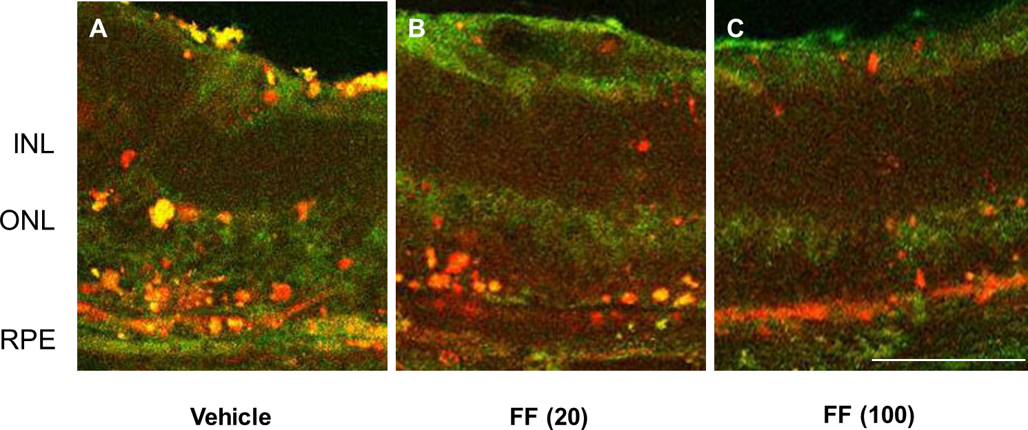
 Figure 3 of
Osada, Mol Vis 2014; 20:1518-1526.
Figure 3 of
Osada, Mol Vis 2014; 20:1518-1526.