Figure 2. Histopathologic features of the retina and histological scores of rats with experimental autoimmune uveoretinitis. The average
histological score of five histological sections from each animal (n=5) was examined. Representative photographs of the groups
treated with vehicle (A), low-dose fenofibrate (FF (20); 20 mg/kg) (B), and high-dose fenofibrate (FF (100); 100 mg/kg) (C) on day 28 post-immunization are shown. The vehicle-treated rats had severe posterior uveoretinitis with destruction of the
photoreceptor cell layer with inflammatory cell infiltration in the retina. Rats treated with low-dose fenofibrate exhibited
mild inflammatory cell infiltration. Rats treated with high-dose fenofibrate exhibited suppression of inflammation and preservation
of the photoreceptor layer. INL; inner nuclear layer, ONL; outer nuclear layer (original magnification, ×100). The mean experimental
autoimmune uveoretinitis (EAU) histological severity grades of the rats treated with vehicle, low-dose fenofibrate, and high-dose
fenofibrate were 4.32±1.65 (n=5), 4.32±1.65 (n=5), and 4.32±1.65 (n=5), respectively. The scores of the rats treated with
high-dose fenofibrate (FF(100)) were significantly lower than that of vehicle- or low-dose fenofibrate-treated rats (D). The results are presented as the mean ± standard deviation (SD) * p<0.0001 versus vehicle-treated group, ** p<0.0001 versus
the low-dose fenofibrate-treated group.
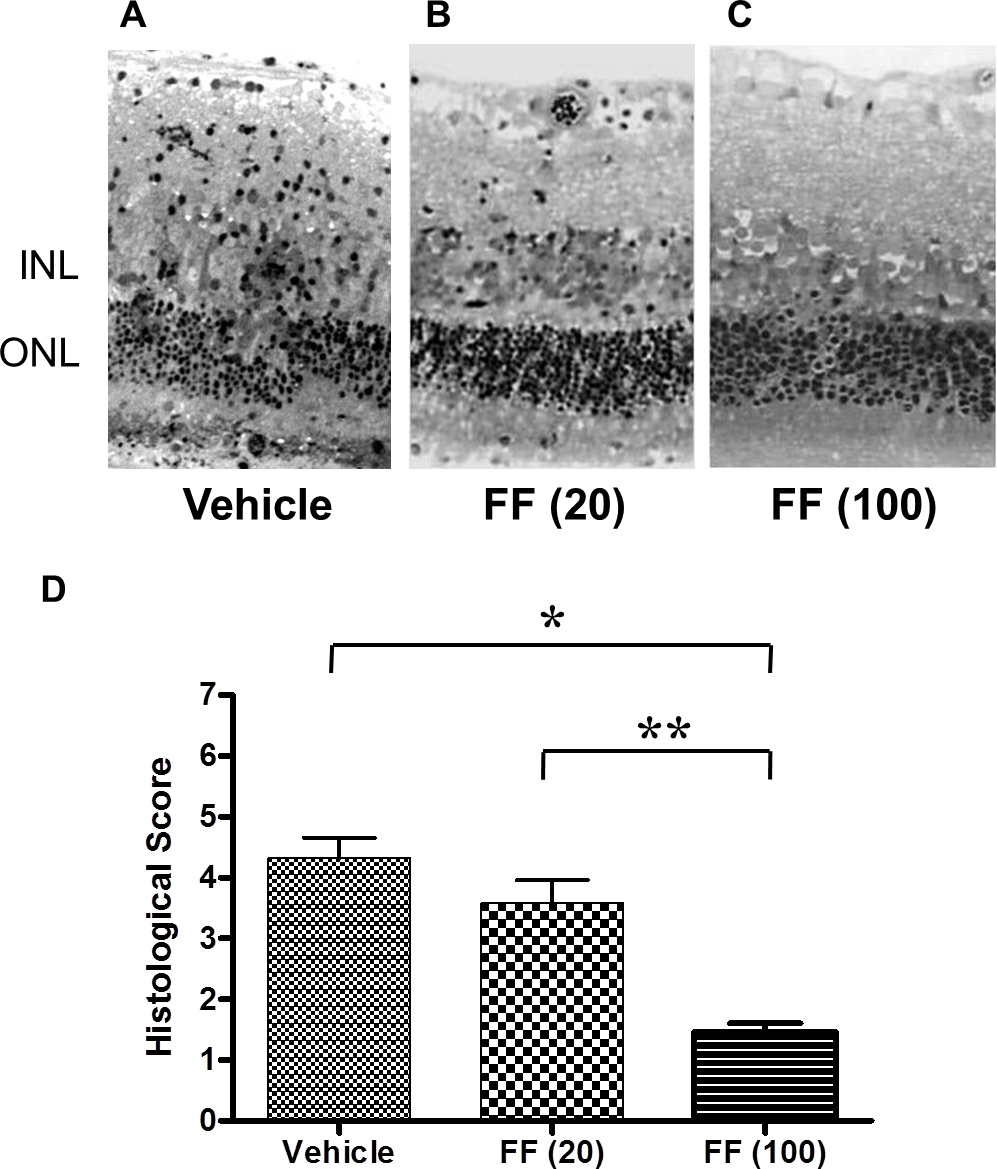
 Figure 2 of
Osada, Mol Vis 2014; 20:1518-1526.
Figure 2 of
Osada, Mol Vis 2014; 20:1518-1526.