Figure 4. Imaging of the oculomotor nerves. A–B: Bilateral oculomotor nerves demonstrated by the axial magnetic resonance imaging (MRI) of a normal control. C–D: The axial MRI indicates bilateral hypoplastic oculomotor nerves in patient II:1. E–F: Patient II:2 presented hypoplasia similar to that of patient II:1, according to the axial MRI.
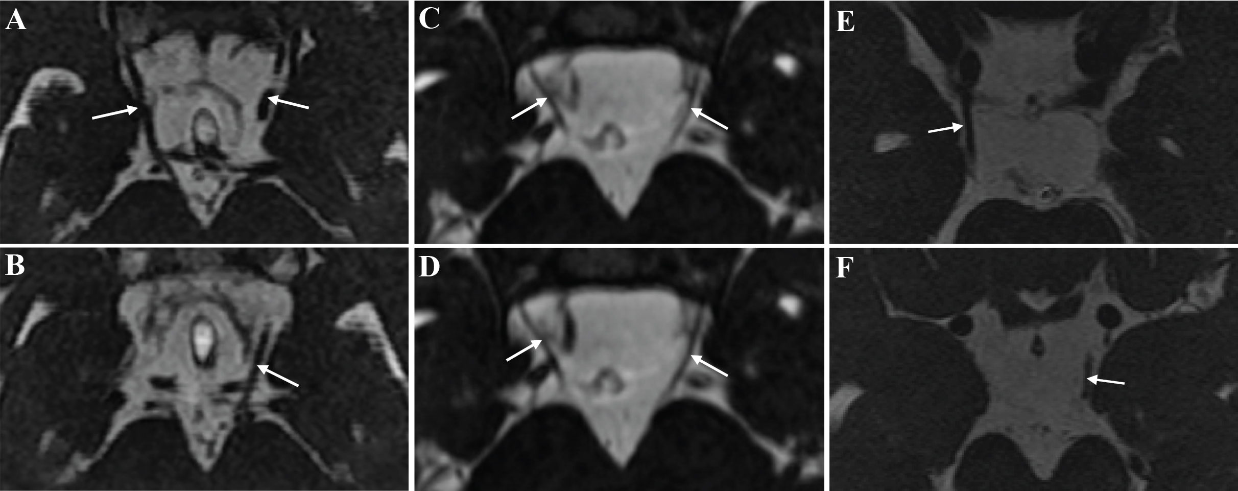
 Figure 4 of
Liu, Mol Vis 2014; 20:15-23.
Figure 4 of
Liu, Mol Vis 2014; 20:15-23.