Figure 3. Imaging of the extraocular muscles and abducens nerves. A–C: Normal aspects of the extraocular muscles (EOMs) and bilateral abducens nerves are presented by coronal and axial magnetic
resonance imaging (MRI) in a control individual. D: Coronal MRI of the bilateral orbits of patient II:1 showed the absence of bilateral superior rectus (SR) muscles and hypoplasia
of the left lateral rectus (LR) muscle but a normal LR muscle of the right eye. E: The axial MRI of patient II:1 showed adduction of the left eye. The LR muscle (arrows) of the left eye was small and had
a string-like configuration, suggesting fibrosis. In contrast, the LR muscle of the right eye was of normal size with a spindle
shape. F: The axial MRI illustrates the absence of the left abducens nerve in patient II:1. G–I: Patient II:2 presented similarly to patient II:1 by showing absence of bilateral SR muscles and small left LR muscle in
the coronal MRI. The axial MRI of both orbits reveals the fibrosis of the left LR muscle (arrows) and adduction of left eye.
An absence of the left abducens nerve was also demonstrated by the axial MRI. Abbreviation: MR, medial rectus; IR, inferior
rectus; SO, superior oblique; ON, optic nerve; BA, basilar artery.
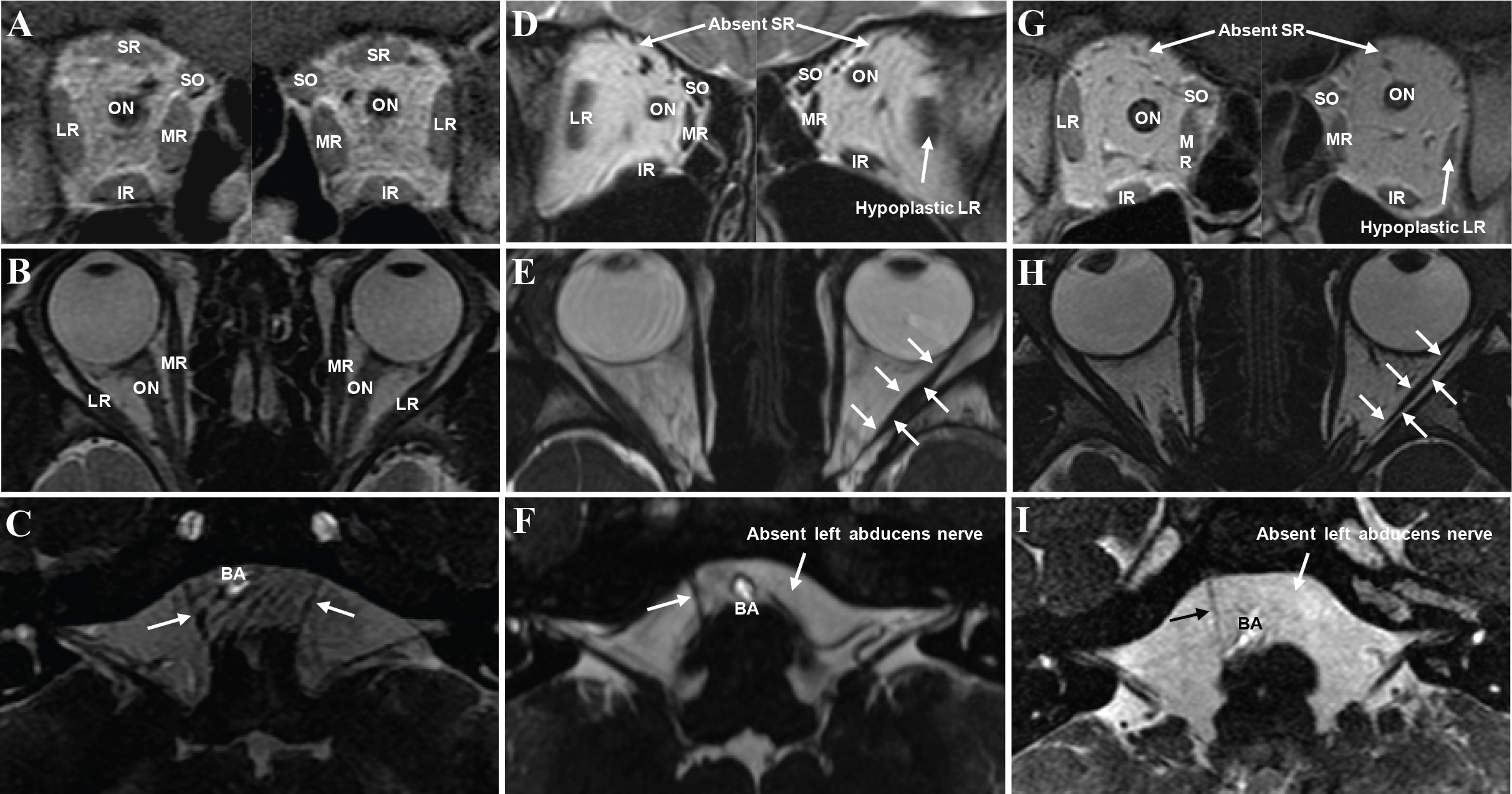
 Figure 3 of
Liu, Mol Vis 2014; 20:15-23.
Figure 3 of
Liu, Mol Vis 2014; 20:15-23.