Figure 2. The absence of Dp71 is associated with histological alterations of the crystalline lens. Cross-sections of crystalline lenses
from wt and KO-Dp71 mice eyes were stained with hematoxylin and eosin at 2 (A-D) and 7 (E-H) months of age. No major alterations in the histology of the crystalline lenses were observed with aging in wt mice (A and E); alternatively, a progressive disorganization was obvious in lenses from KO-Dp71 mice (B and F). Higher magnification images suggest alterations in the ultrastructure of the lenses in KO-Dp71 mice (D and H, white arrows) along with a disorganized transition zone at both 2 and 7 months of age (C versus D and G versus H, respectively; black arrows; e: epithelial cells; f: fiber cells; tz: transition zone). Scale bars A, B, E, and F: 300 μm; Scale bars C, D, G, and H: 80 μm.
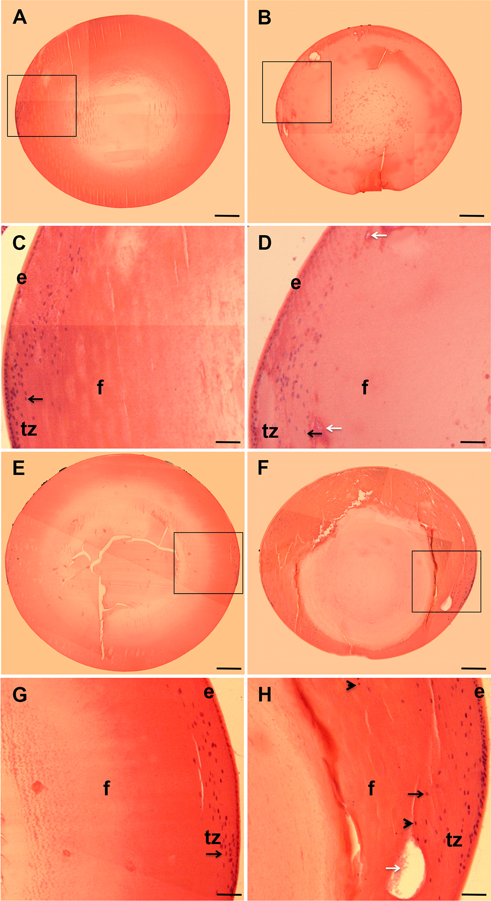
 Figure 2 of
Fort, Mol Vis 2014; 20:1480-1490.
Figure 2 of
Fort, Mol Vis 2014; 20:1480-1490.