Figure 2. For all panels, retinal Müller cells were grown in normal glucose (NG) or high glucose (HG) and transfected with an IRS-1
plasmid or a mutated IRS-1 plasmid and treated with TNF-α or TNF-α+mutant (Panels A–B) with retinal cell lysates used for all experiments. A: The control demonstrates successful transfection of the normal IRS-1 plasmid and mutant IRS-1 plasmid. B: The ELISA results for decreased phosphorylation of IRS-1 on serine 307 when TNF-α is added with the mutant. *p<0.05 versus
NG non-treated; #p<0.05 versus HG non-treated; $p<0.05 versus HG TNF-α. n=5 for all groups. Data are mean±standard error of
the mean (SEM).
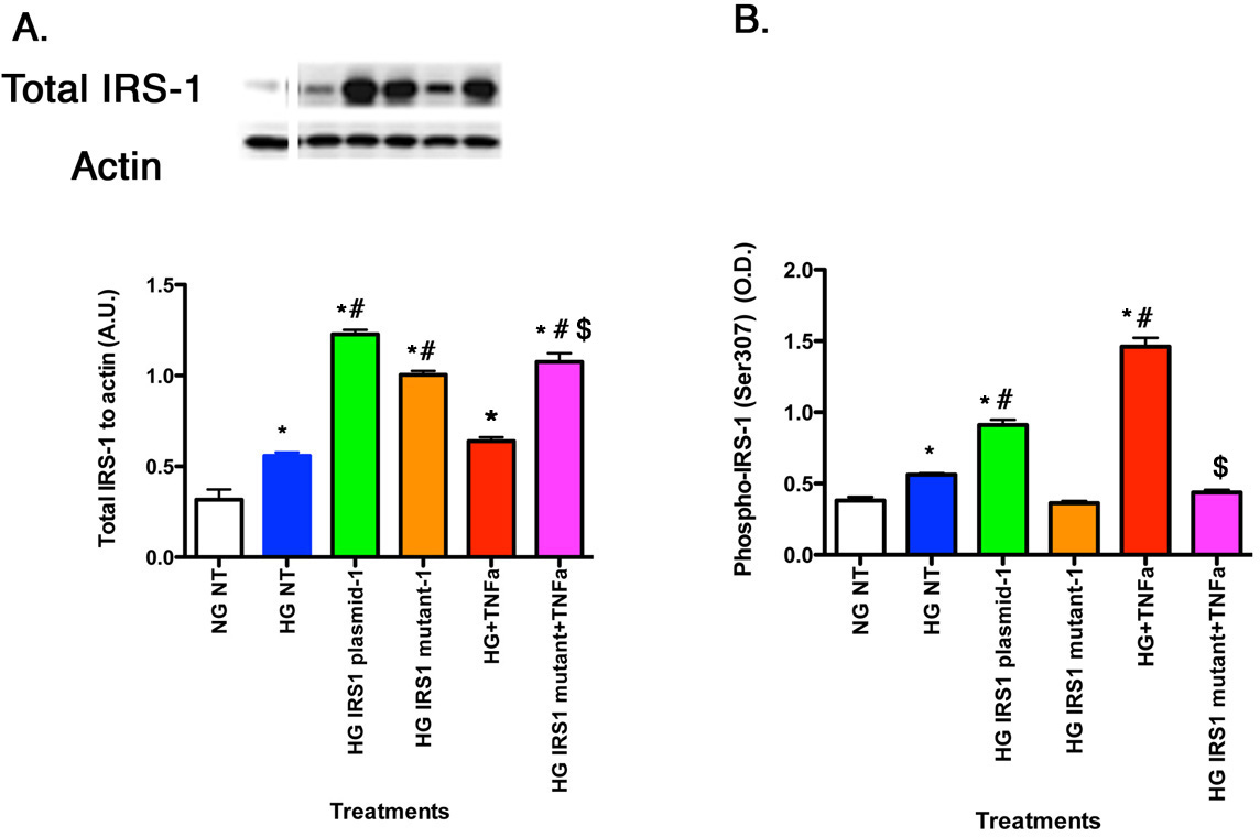
 Figure 2 of
Jiang, Mol Vis 2014; 20:1463-1470.
Figure 2 of
Jiang, Mol Vis 2014; 20:1463-1470.