Figure 3. Neurite outgrowth from retinal ganglion cells (RGCs) was investigated by immunoreactivity to α-tubulin. Oxidative stress was
induced by exposure to 20 μM of H2O2 for 16 h. In the maltol treatment group, 1 mM of maltol was added to the culture medium at the time of injury. A: Undamaged control; B: Oxidative stress only; C: Oxidative stress with maltol treatment. Neurites were labeled with green fluorescence and nuclei were counterstained with
blue fluorescence. Scale bar is 25 μm. D: The proportion of cells having neurites and interacting with surrounding cells was expressed as mean ± standard error of
the mean (SEM). Asterisks, p<0.05, n=12, *Significant difference between control & oxidative stress with maltol treatment
vs. oxidative stress only, **Significant difference between control & oxidative stress only vs. oxidative stress with maltol
treatment.
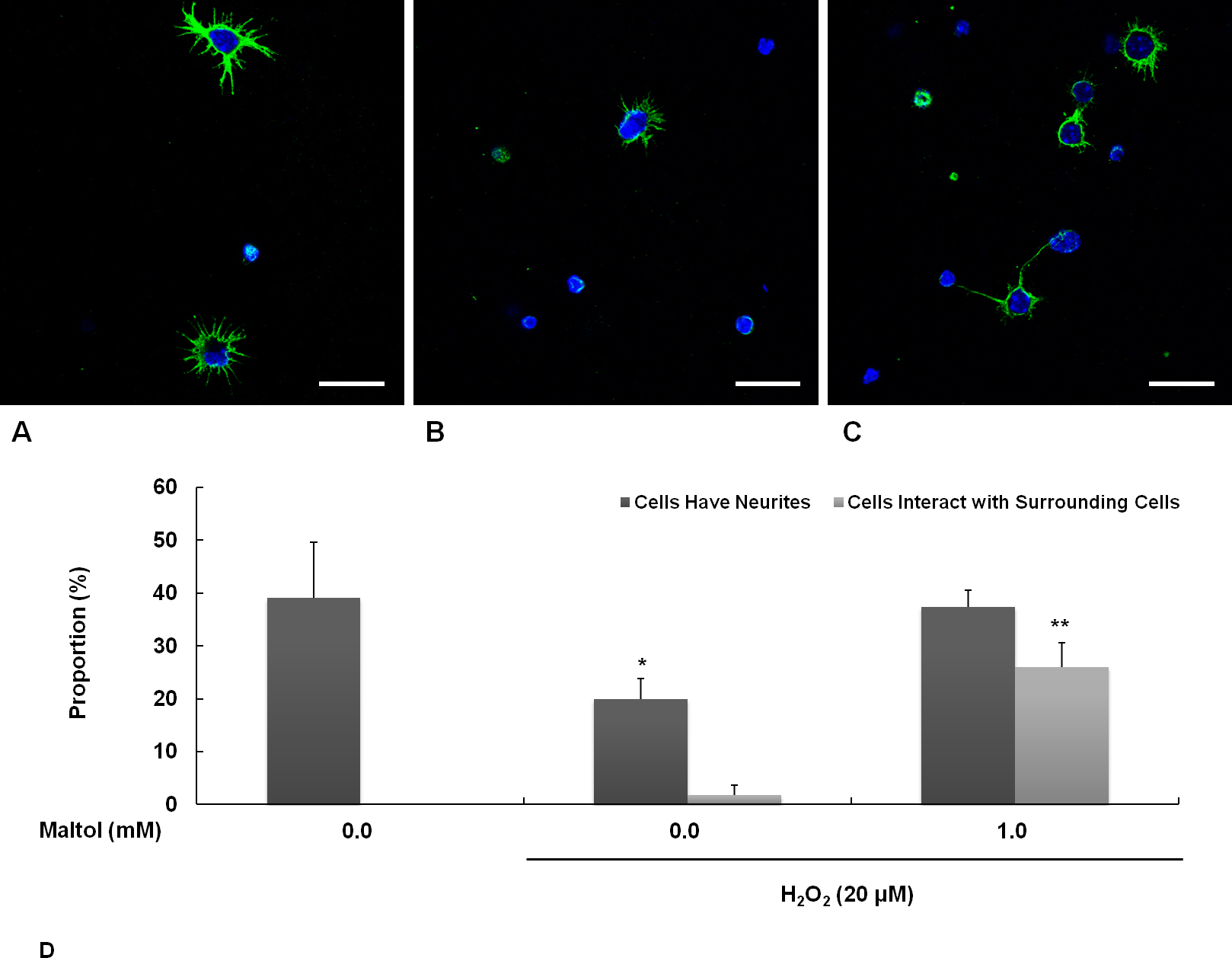
 Figure 3 of
Hong, Mol Vis 2014; 20:1456-1462.
Figure 3 of
Hong, Mol Vis 2014; 20:1456-1462.