Figure 2. Apoptosis was evaluated by terminal deoxynucleotidyl transferase (TdT)-mediated deoxyuridine triphosphate (dUTP) nick end
labeling (TUNEL). Oxidative stress was induced by exposing the retinal ganglion cells (RGCs) to 20 μM of H2O2 for 16 h. In the maltol treatment group, 1 mM of maltol was added to culture medium at the time of injury. A: Undamaged control; B: Oxidative stress only; C: Oxidative stress with maltol treatment. Apoptotic cells showed broken nuclei with green fluorescence over counterstained,
unfragmented nuclei with orange-red fluorescence. Scale bar is 200 μm. D: The proportion of apoptotic cells was expressed as mean ± standard error of the mean (SEM). Asterisks, p<0.05, n=12, *Significant
difference between control versus oxidative stress with/without maltol treatment, **Significant difference between oxidative
stress without versus with maltol treatment.
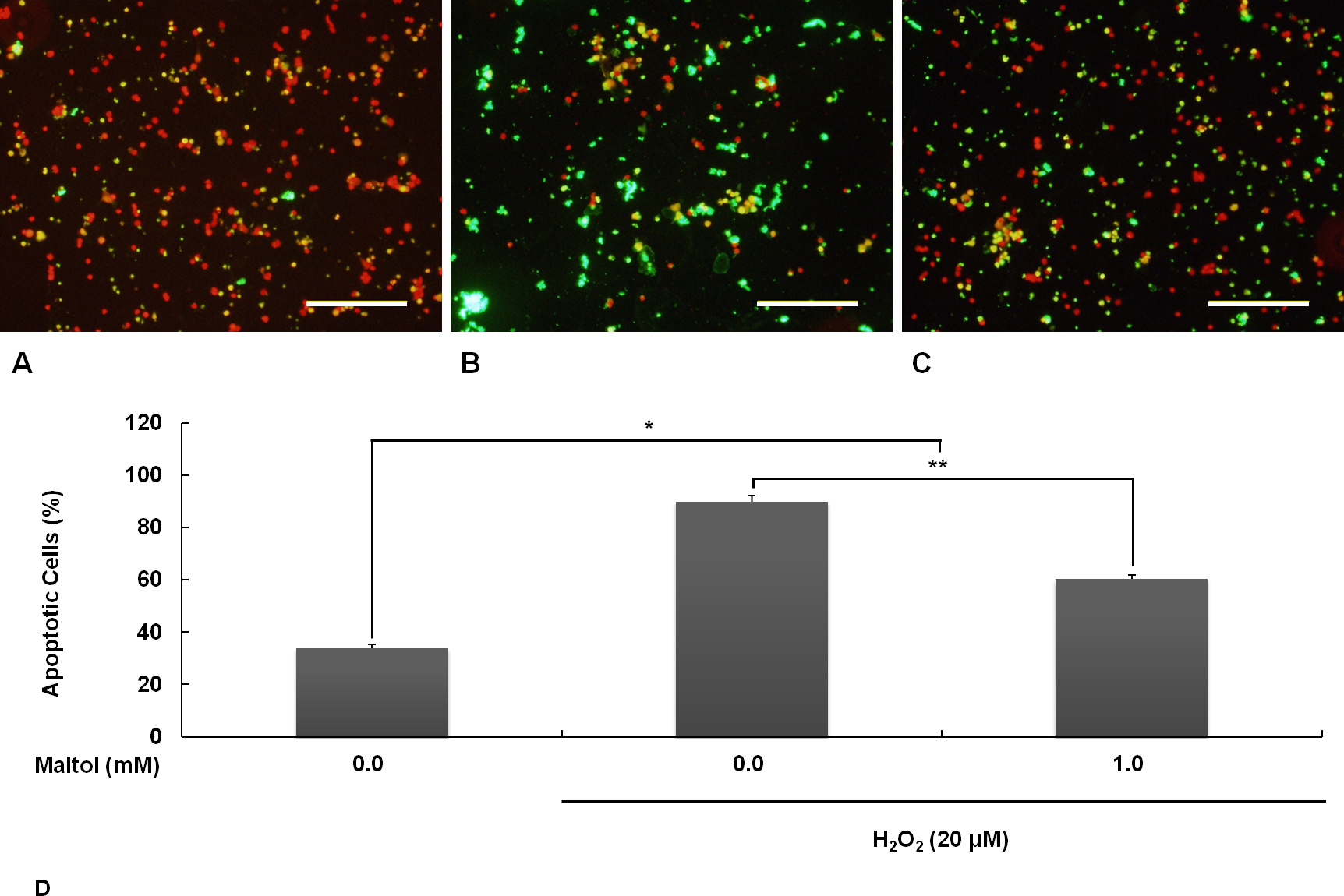
 Figure 2 of
Hong, Mol Vis 2014; 20:1456-1462.
Figure 2 of
Hong, Mol Vis 2014; 20:1456-1462.