Figure 7. Mas expression in retinal vessels and cultured cell lines. A: The Mas receptor protein detected with immunofluorescence and colocalization with endothelial cell marker (CD31) in trypsin
digested retinal vasculature preparation. B–E: Mas expression detected with immunofluorescence in (B) human Müller cells, (C) mouse N9 microglial cells, (D) human retinal pigment epithelial (RPE, ARPE19) cells, and (E) cone photoreceptor cells (661W). Nuclei were counterstained with 4',6-diamidino-2-phenylindole (DAPI). B–E: Inserts show Mas expression alone. Scale bar=20 μm.
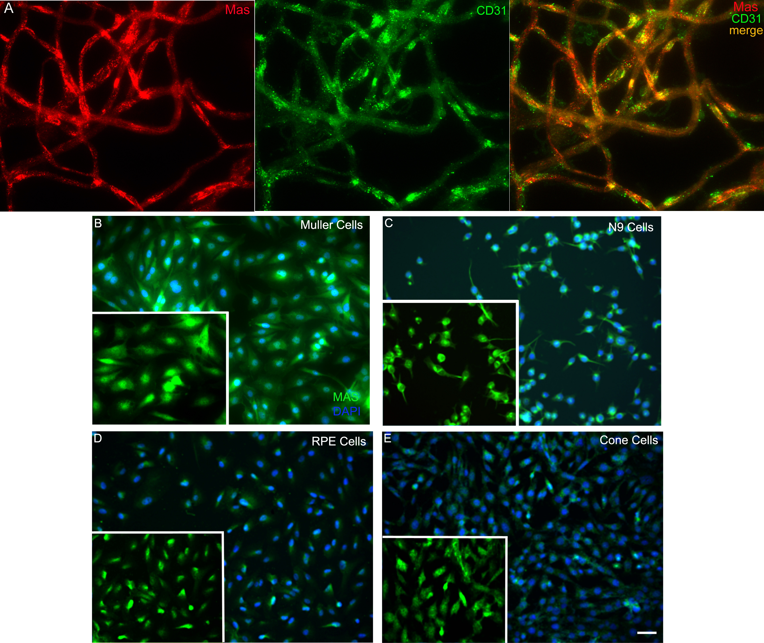
 Figure 7 of
Prasad, Mol Vis 2014; 20:1443-1455.
Figure 7 of
Prasad, Mol Vis 2014; 20:1443-1455.