Figure 6. In situ detection of Mas receptor mRNA expression in developing and adult mouse retinas. A–F: Mas receptor mRNA was detected with in situ hybridization in the retinas of different ages (P1–P60). D’–F’: Lower magnification (100X) image showing Mas expression in the RGC layer at P15, P21, and P60. G: Positive control: In situ detection of peptidylprolyl isomerase B (PIPB) in the P60 mouse retina. H: Positive control: In situ detection of arrestin in the P60 mouse retina. I: Negative control in the P60 mouse retina. NBL=neuroblast layer; ONL: outer nuclear layer; OPL=outer plexiform layer; INL=inner
nuclear layer; IPL=inner plexiform layer; RGC=retinal ganglion cell layer. Scale bar=50 μm (A–D); D’–F’=10 μm.
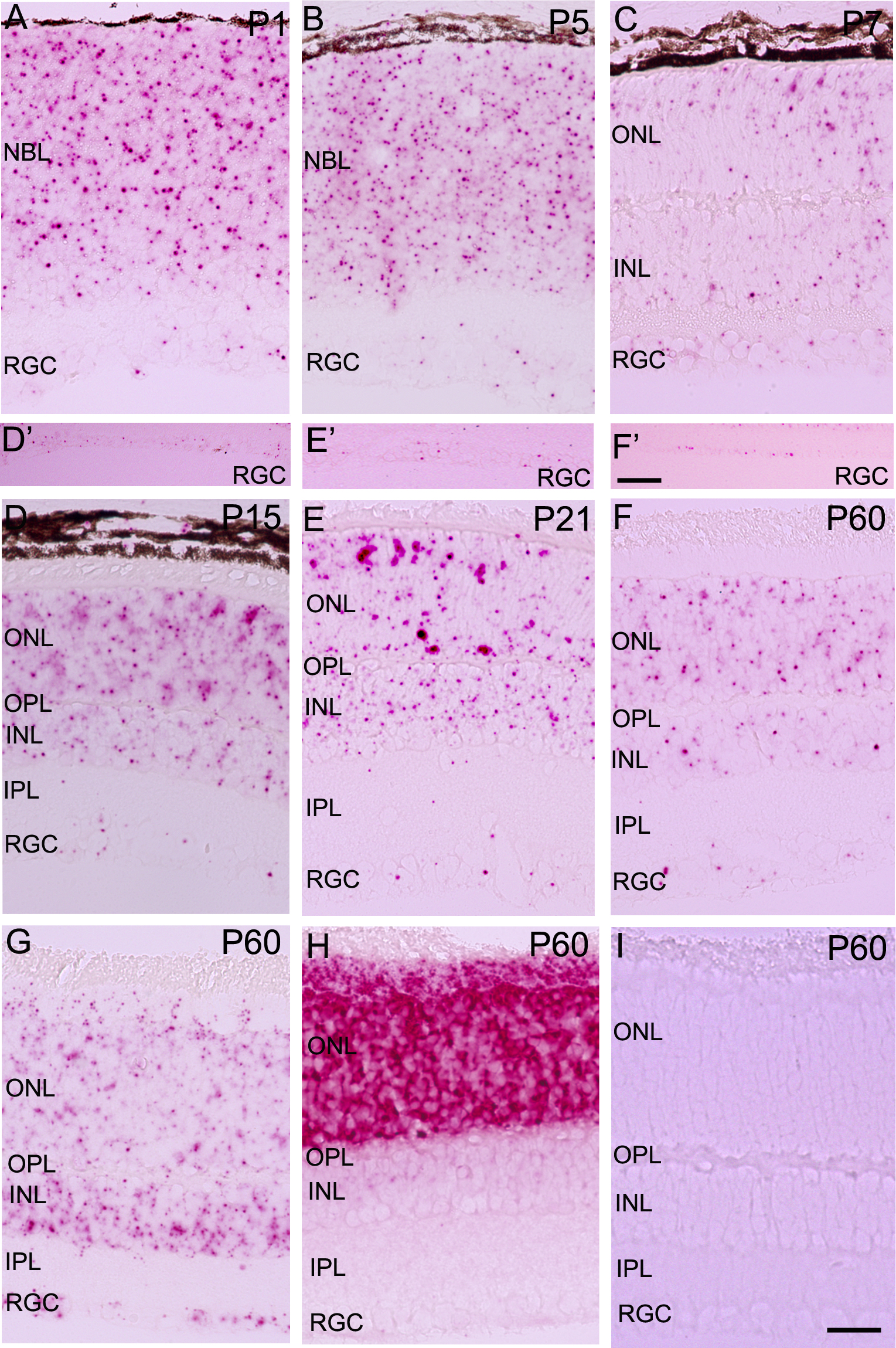
 Figure 6 of
Prasad, Mol Vis 2014; 20:1443-1455.
Figure 6 of
Prasad, Mol Vis 2014; 20:1443-1455.