Figure 4. Cellular localization of the Mas receptor protein in the RPE and the ciliary body of the mouse eye. Panel A-C: Double immunostaining for the RPE cell marker (RPE 65, green) and Mas (red) in the CD1 mouse retina. D: Negative control for RPE65 and Mas staining in the RPE cell layer. E and F: Immunofluorescence detection of Mas in the ciliary body of the CD1 mouse eye. G: Negative control for Mas staining in the ciliary body of the CD1 mice. RPE=retinal pigmented epithelium; CC=choroidal capillaries.
Scale bar=20 μm (A-D); 50 μm (E-G).
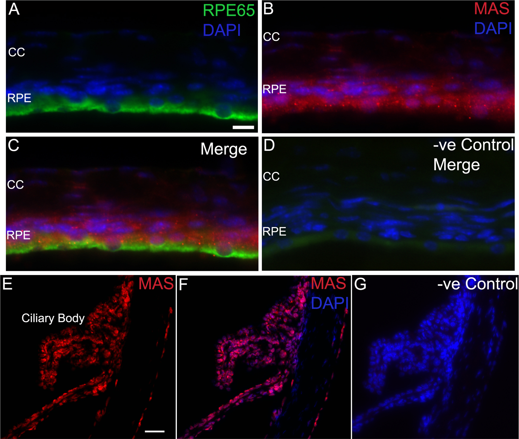
 Figure 4 of
Prasad, Mol Vis 2014; 20:1443-1455.
Figure 4 of
Prasad, Mol Vis 2014; 20:1443-1455.