Figure 3. Cellular localization of the Mas receptor protein in retinal glial cells. Double immunostaining for glial cell markers and
the Mas receptor in the mouse retina shows a low level expression of Mas in (A-C) astrocytes which are glial fibrillary acidic protein (GFAP) positive and in (D-I) Müller cell bodies that are p27KIP1 and CRALBP positive. The nuclei were counterstained with 4',6-diamidino-2-phenylindole (DAPI). INL=inner nuclear layer; RGC=retinal
ganglion cell layer. Scale bar=50 µm (A-C); 20 µm (D-I).
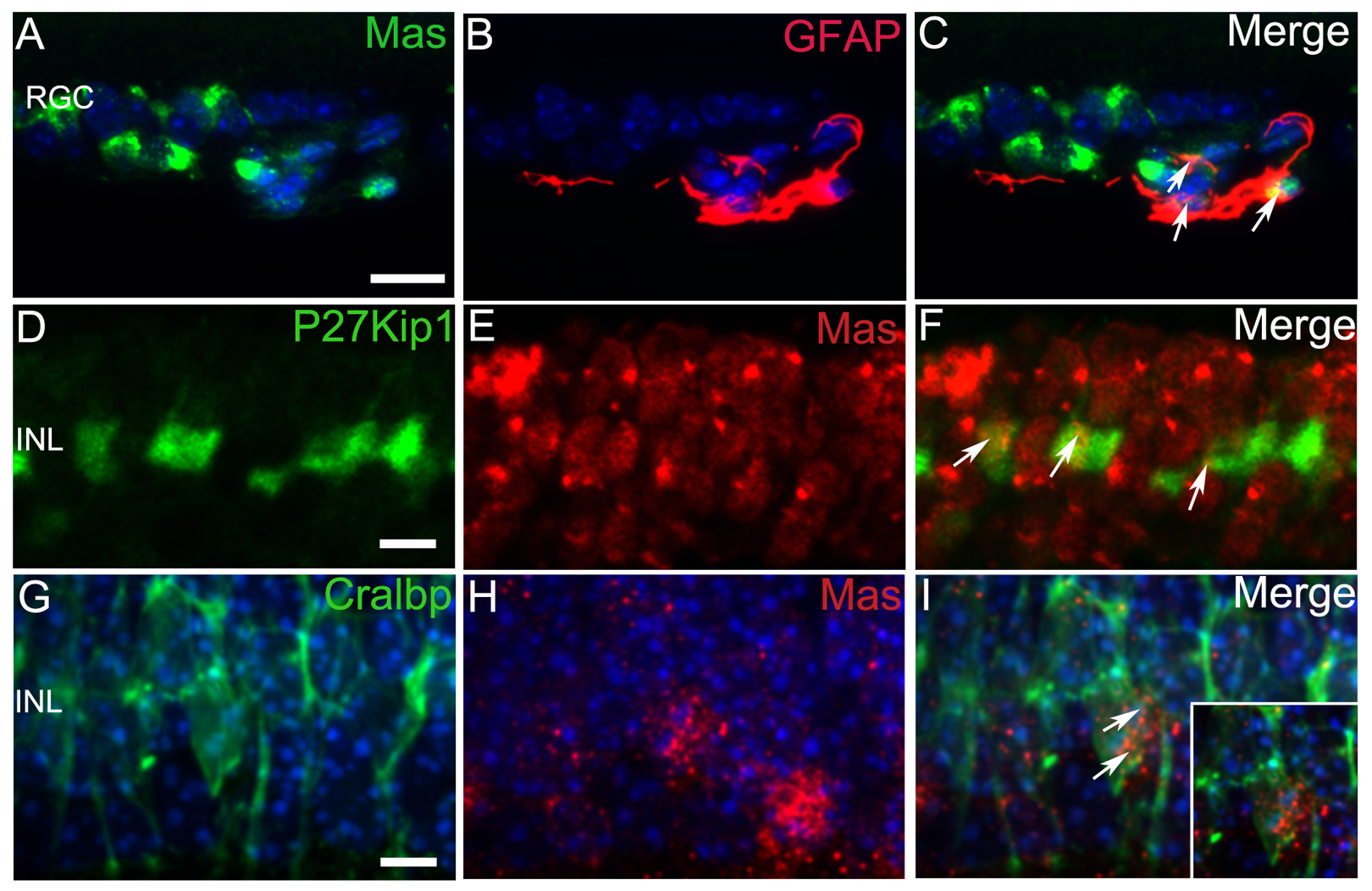
 Figure 3 of
Prasad, Mol Vis 2014; 20:1443-1455.
Figure 3 of
Prasad, Mol Vis 2014; 20:1443-1455.