Figure 7. Model of DSCAM dynamic localization. A: At early developmental time points, DSCAM was localized along the developing neurites. As development proceeded, DSCAM took
on a punctate pattern and overlapped with synaptic markers at time points consistent with synaptogenesis, but after ectopic
adhesions had developed. As the retina matured, DSCAM was not observed overlapping with synapses but was observed overlapping
with catenins. B: DSCAM prevented adhesion, but in the absence of the protein, other factors, possibly cadherins or other CAMs such as sidekicks,
caused similar cells and their neurites to adhere to each other.
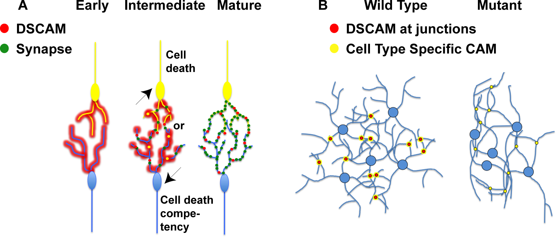
 Figure 7 of
Belem de Andrade, Mol Vis 2014; 20:1422-1433.
Figure 7 of
Belem de Andrade, Mol Vis 2014; 20:1422-1433.