Figure 6. Synaptic and adhesion molecules in the wild-type and Dscam mutant retina. Wild-type and Dscam mutant retina sections were stained with antibodies to synaptic or junction molecules. A and B: Section of wild-type and Dscam mutant retina were stained with antibodies to CTBP2. C and D: Sections of wild-type and Dscam mutant retina were stained with antibodies to PSD95. E and F: Sections of wild-type and Dscam mutant retina were stained with antibodies to Bassoon. G and H: Sections of wild-type and Dscam mutant retina were stained with antibodies to α-n-catenin. I and J: Sections of wild-type and Dscam mutant retina were stained with antibodies to GABAaδ2. K and L: Sections of wild-type and Dscam mutant retina were stained with antibodies to γ-protocadherin. A similar distribution of synaptic markers, considering the
disorganization of the Dscam mutant retina, was observed in the genotypes. Scale = 53.25 μm.
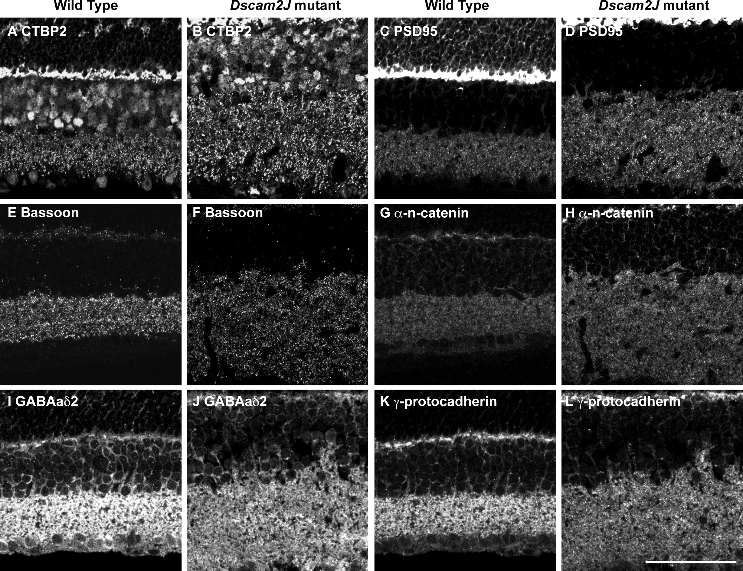
 Figure 6 of
Belem de Andrade, Mol Vis 2014; 20:1422-1433.
Figure 6 of
Belem de Andrade, Mol Vis 2014; 20:1422-1433.