Figure 5. Developmental colocalization between DSCAM and synaptic and scaffolding molecules. A–D: Retina sections at early postnatal stages were stained with antibodies to DSCAM and Sap102 or GluR2 or GABAaδ2 or γ-protocadherin.
Flipped controls were generated by rotating one of the channels 180 degrees across the vertical axis (to preserve S1–S5 but
provide a randomized control). The use of flipped controls maintained the degree of immunoreactivity and provided a measure
of incidental colocalization. Compared to the flipped controls, an increase in colocalization of all antigens and DSCAM was
observed. E: Section of the P0 retina stained with antibodies to DSCAM and γ-protocadherin. Both proteins overlapped in a diffuse pattern
across the inner plexiform layer. F: Primary culture of retinal ganglion cells stained with antibodies to DSCAM and γ-protocadherin. Overlap was observed in
some but not all puncta (yellow arrows; overlap, red or green arrow; no overlap). The scale bar in (F) is equivalent to 53.25 µm in A–D (insets are 7.9 µm), 100 μm in E, and 5.17 μm in F.
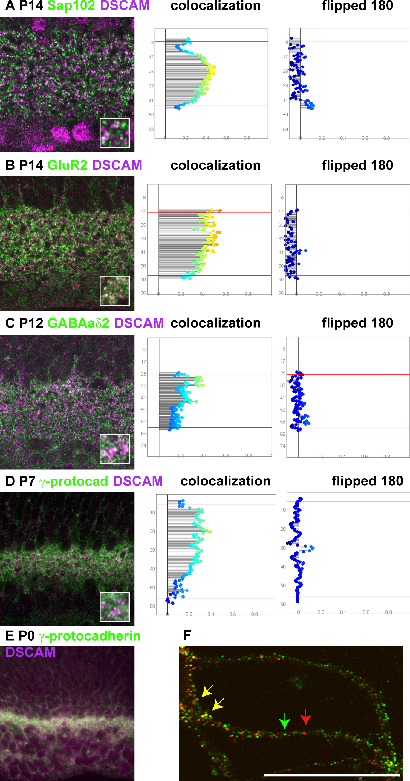
 Figure 5 of
Belem de Andrade, Mol Vis 2014; 20:1422-1433.
Figure 5 of
Belem de Andrade, Mol Vis 2014; 20:1422-1433.