Figure 4. Robust wave of developmental colocalization between DSCAM and PSD95. A–E: Sections of the P4, P7, P10, P14, and adult retina were stained with antibodies to DSCAM and PSD95. Colocalization was plotted
regarding the laminar depth and as a volcano plot. Flipped controls were generated by rotating one of the channels 180 degrees
across the vertical axis (to preserve S1–S5 but provide a randomized control). The use of the flipped controls maintained
the degree of immunoreactivity and provided a measure of incidental colocalization. Limited colocalization between DSCAM and
PSD95 was observed at P4 (A) and P7 (B) and in the adult retina (E). At P10 and P14, many instances of overlap between PSD95 and DSCAM were observed (C and D) while colocalization analysis identified a higher degree of colocalization compared to flipped controls or earlier and later
time points. The scale bar in E is equivalent to 53.25 µm in A–E and 7.9 µm in insets from A–E.
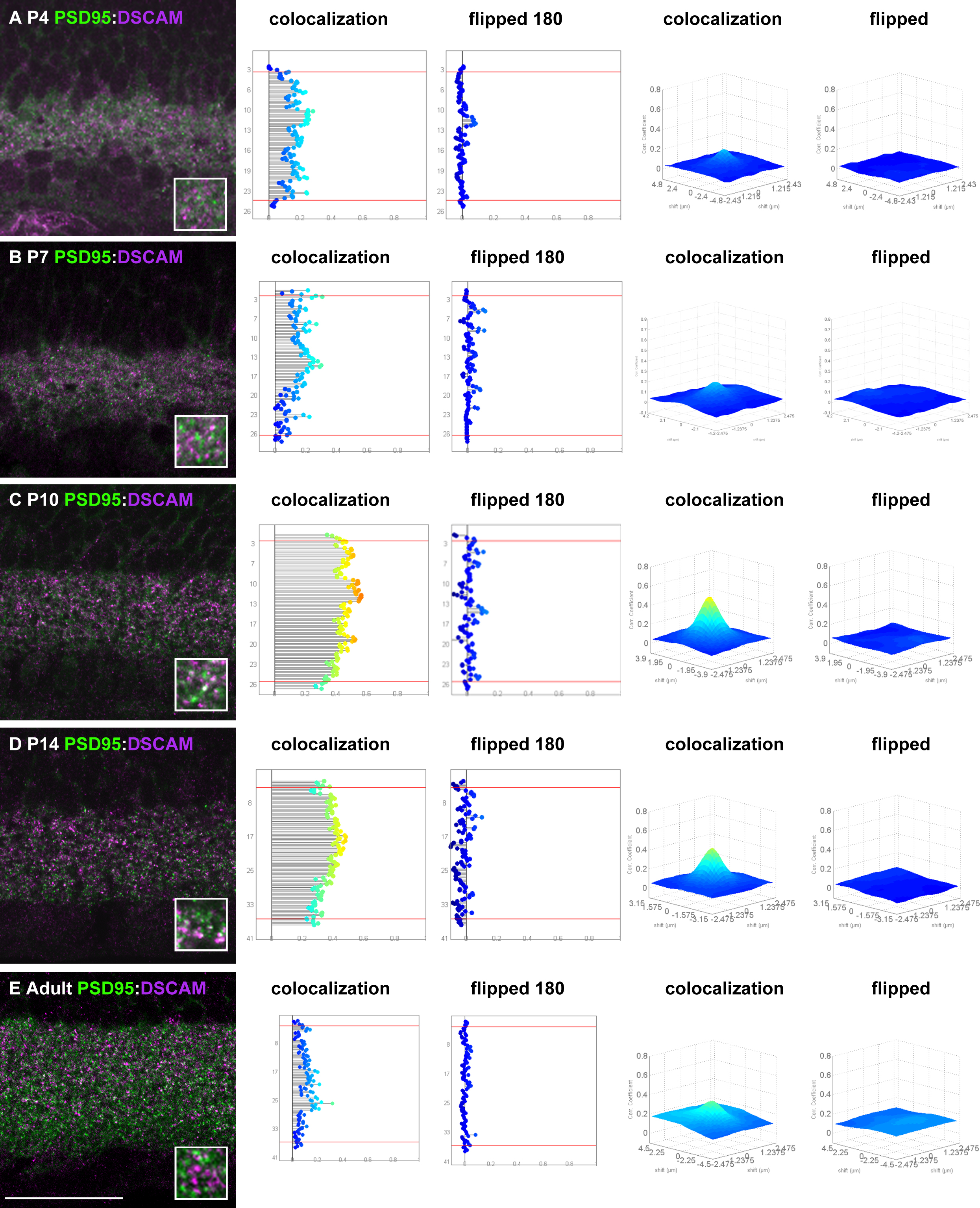
 Figure 4 of
Belem de Andrade, Mol Vis 2014; 20:1422-1433.
Figure 4 of
Belem de Andrade, Mol Vis 2014; 20:1422-1433.