Figure 3. Limited colocalization of DSCAM and synaptic markers detected in the adult retina. Analysis of colocalization between DSCAM
and C-Terminal Binding Protein-2 (CTBP2; A), NMDA R1 (NR1) (B), Synapsin (C), Neuroligin (D) NMDA 2B (E), NMDA 2AB (F) Synapse Associated Protein 102 (SAP102; G), Shank (H), P2X2 (I), P2X7 (J), Glutamate Receptor 4 (GluR4; K), GABAaδ2 (L), Calcium/Calmodulin-Dependent Serine Protein Kinase (CASK; M), Bassoon (N), Vesiclar Glutamate Transporter 3 (vglut3; O), Syntaxin 2 (P), RIM 2 (Q), Glycine Receptor (R), Glutamate Receptor 1 (GluR1; S), Oxidative Phosphorylation marker (T), Membrane Associated Guanylate Kinase 2 (MAGI2; U), Connexin 36 (V), 14-3-3 ε (W), Syntaxin 4 (X), ZO-1-Associated Nucleic Acid-Binding Protein (ZONAB; Y), Membrane Associated Guanylate Kinase 3 (MAGI3; Z), Zonula Occludens-2 (AA) or 14-3-3 ξ (BB). CC and DD, Dispersed cultures of retinal ganglion cells were stained with antibodies to DSCAM, PSD95 and GABAa∂2. Only rare examples
of DSCAM overlap with synaptic markers were observed (CC arrow). The scale bar in DD is equivalent to 53.25 µm in A - CC and 7.9 µm in insets from A - CC. The scale bar in DD is equivalent to 15.8 µm in CC and 35.5 µm in DD.
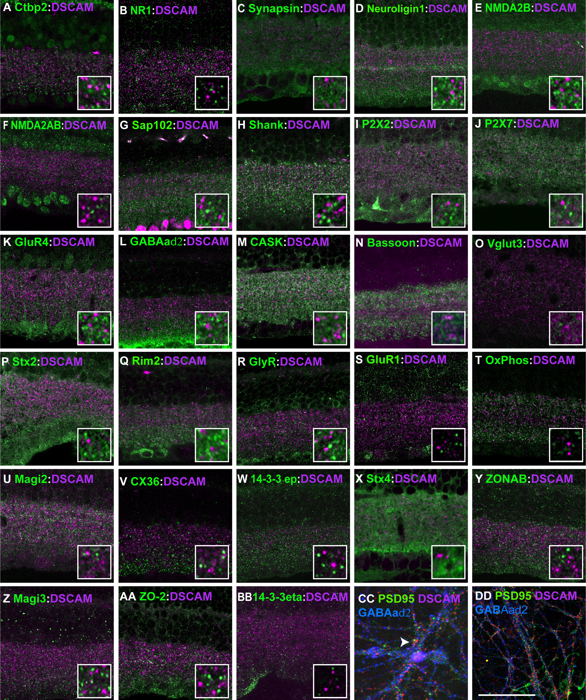
 Figure 3 of
Belem de Andrade, Mol Vis 2014; 20:1422-1433.
Figure 3 of
Belem de Andrade, Mol Vis 2014; 20:1422-1433.