Figure 2. DSCAM colocalizes with catenins in the adult retina. A-C: Sections of retina were stained with antibodies to DSCAM, PSD95 and bassoon. DSCAM staining appeared to overlap minimally
with synaptic markers. D: Retinal sections were stained with two different antibodies to DSCAM. Colocalization of markers was apparent in white (inset
box). A bar chart readout of the colocalization by laminar depth, running from S1 (top) to S5 (bottom). The degree of protein
colocalizing is indicated by the length of bar on the x-axis. Flipped controls were generated by rotating one of the channels
180 degrees across the vertical axis (so as to preserve S1-S5 but provide a randomized control). E: Retinal sections were stained with antibodies to bassoon and GABAa∂2. Colocalization of markers was apparent in white and
in the bar chart (inset box). F: Retinal sections were stained with antibodies to GluR2 and bassoon. No colocalization of markers was apparent in white
(inset box), and as depicted in the bar chart. G, Retinal sections from adult mice were stained with antibodies to Syntaxin1 and DSCAM. Minimal colocalization of markers
was apparent (inset box) and in the bar charts. H: Retinal sections from adult mice were stained with antibodies to Syntaxin3 and DSCAM. No colocalization of markers was apparent
in white (inset box) and as depicted in the bar charts. I: Retinal sections from adult mice were stained with antibodies to the adherens junctions marker ß-catenin and DSCAM. Colocalization
of markers was apparent in white (inset box) and in the bar charts. J: Retinal sections from adult mice were stained with antibodies to the adherens junctions marker α-n-catenin and DSCAM. Colocalization
of markers was apparent in white (inset box) and in the bar charts. K: Retinal sections from adult mice were stained with antibodies to the junction marker Zonula Occludens-1 and DSCAM. No colocalization
of markers was apparent in white (inset box) or as depicted by the bar charts. L: Retinal sections from adult mice were stained with antibodies to the gap junction marker Connexin 45 and DSCAM. No colocalization
of markers was apparent in white (inset box) or by the bar charts. The scale bar in (A) is equivalent to 12.5 µm in A, 18.9 µm in B and C, 53.25 µm in D-L and 7.9 µm in the insets from D-L.
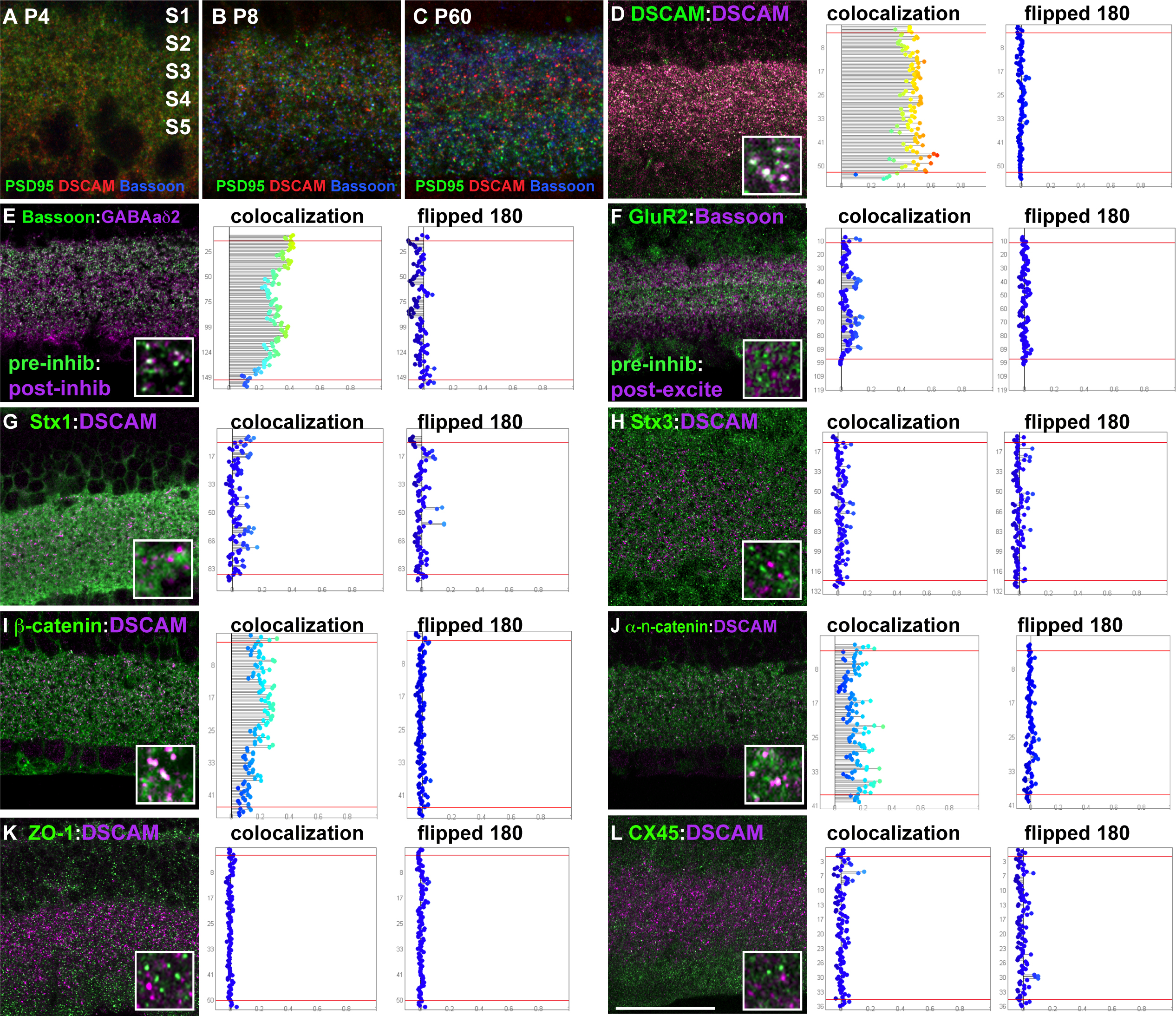
 Figure 2 of
Belem de Andrade, Mol Vis 2014; 20:1422-1433.
Figure 2 of
Belem de Andrade, Mol Vis 2014; 20:1422-1433.