Figure 1. Localization of DSCAM in the mouse retina. A–D: Sections of wild-type retina from P0, P2, P4, and P7 mice were stained with antibodies to DSCAM. The DSCAM protein was observed
throughout the inner plexiform layer (IPL) at P0 and P2. By P4, and especially by P7, DSCAM immunoreactivity was more punctate
compared to earlier ages. E: DSCAM was observed throughout the IPL and in a limited fashion in the outer plexiform layer (OPL) of the adult retina. F: Residual DSCAM protein in the Dscamdel17 allele accumulated around the cell soma. G: The DSCAM protein was not observed in the Dscam2J allele. H and I: The DSCAM protein in the Dscam3J and DscamFD alleles aggregated around the cell soma. J: A secondary antibody-only control demonstrated the lack of nonspecific labeling by the secondary antibody. K–O: Sections of the wild-type and Dscamdel17 retina were stained with hematoxylin and eosin. Disorganization of the Dscamdel17 retina became apparent by approximately P4, when cells were observed in the inner plexiform layer (arrows) and the retinal
ganglion cell layer was thicker compared to wild-type (G; arrows) and was easily observable by P6, when uneven lamination of the retinal ganglion and inner nuclear layer was observed
(H; arrows). The scale bar in (I) is equivalent to 134 µm in A–J, 91 µm in K, 116 µm in L, 170 µm in M and N, and 156 µm in O. The insets in B and C are 3.2 µm wide.
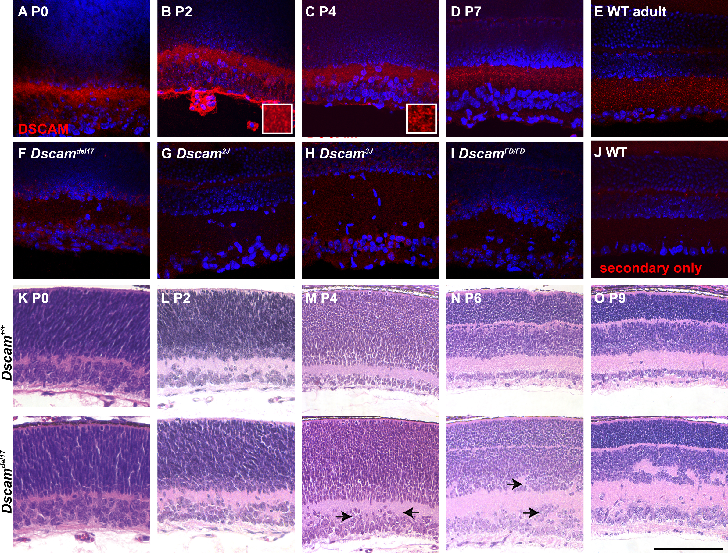
 Figure 1 of
Belem de Andrade, Mol Vis 2014; 20:1422-1433.
Figure 1 of
Belem de Andrade, Mol Vis 2014; 20:1422-1433.