Figure 4. 5-aza-2’-deoxycytidine (5-aza-dC) and trichostatin A (TSA) induced pigment epithelium derived factor (PEDF), reverses PEDF/
vascular endothelial growth factor (VEGF) ratio and mitigated the up-regulations of exacerbating factors at protein levels
in human retinal endothelial cells (HRECs) and human retinal pigment epithelial (HRPE) cells in high glucose (HG) or interleukin
(IL)-1β condition while TSA further up-regulated intercellular cell adhesion molecule-1 (ICAM-1) protein in HRECs. Cells were
cultured for 24 h (IL-1β stimulation) or 48 h (HG stimulation) with or without 5-aza-dC (5 μM, 10 μM) or TSA (0.2 μM, 0.5
μM, 1 μM). PEDF, VEGF, ICAM-1, IL-1β, and matrix metalloproteinase 2 (MMP2) secretions and the PEDF/VEGF protein ratio were
analyzed with enzyme-linked immunosorbent assay (ELISA). The data are expressed as the mean ± standard deviation (SD). The
expression was normalized separately to its control counterpart. *p<0.05 versus the normal physiologic glucose (NG) group,
#p<0.05 versus the HG group or the IL-1β group. All experiments were repeated three times with similar results. IL-1β secretion
could not be detected in this study. (IL-1β secretion could not be analyzed due to due to its low concentration [the HG group]
or the exogenous human recombination IL-1β [the IL-1β stimulation group])”.
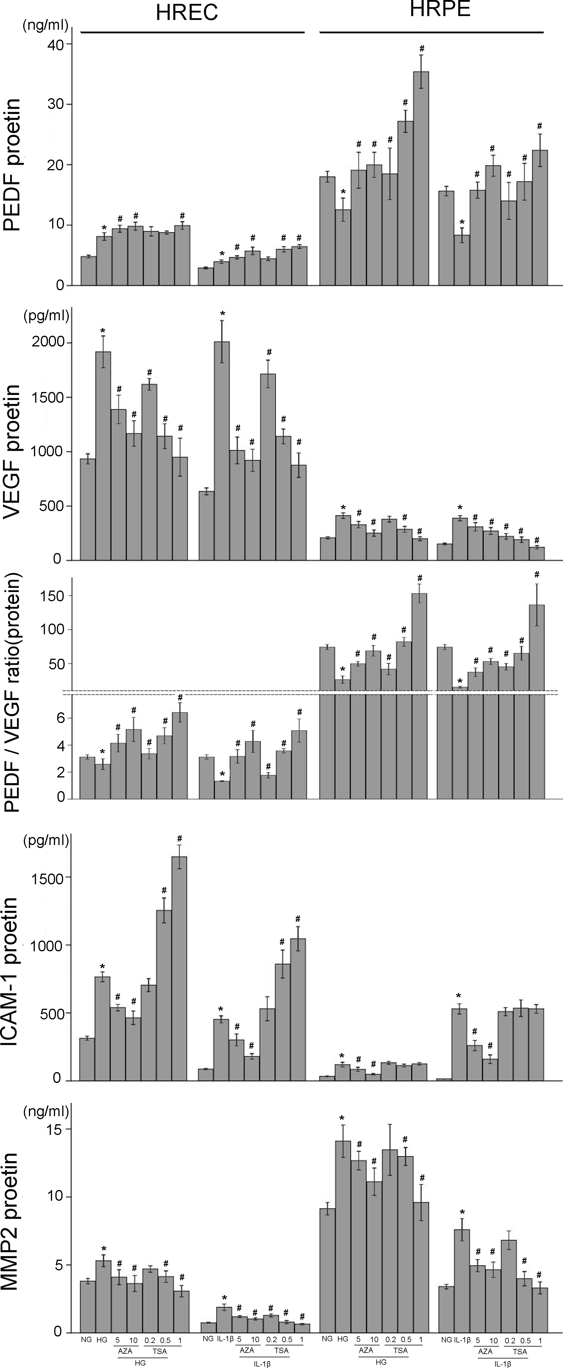
 Figure 4 of
Xie, Mol Vis 2014; 20:1411-1421.
Figure 4 of
Xie, Mol Vis 2014; 20:1411-1421.