Figure 3 of
García-García, Mol Vis 2014; 20:1398-1410.
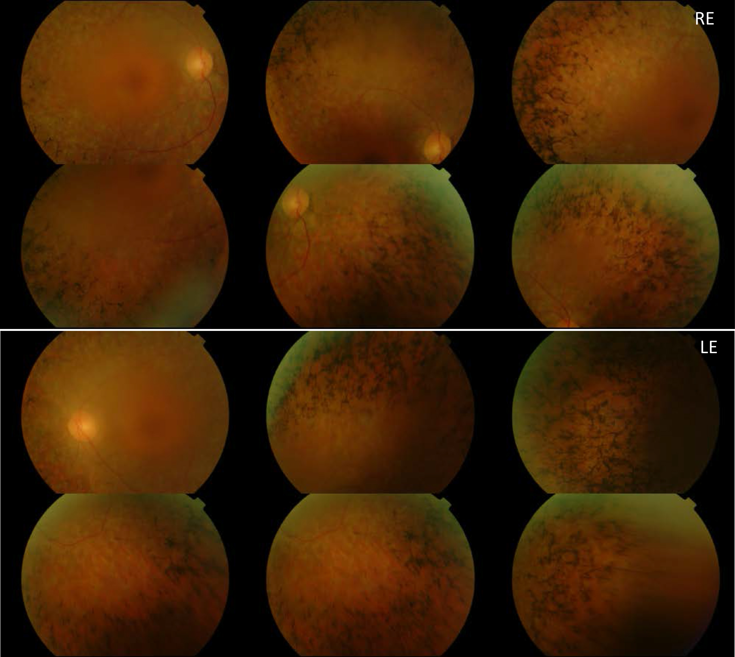
Figure 3.
Eye fundus images obtained after examination of patient RP-1397 show bone spicule deposits, attenuation of vessels and waxy pallor of the optic nerve head in both eyes.
 Figure 3 of
García-García, Mol Vis 2014; 20:1398-1410.
Figure 3 of
García-García, Mol Vis 2014; 20:1398-1410.