Figure 6. Hepatitis C virus core and NS3 induced apoptosis and cell death. Human conjunctival fibroblasts were exposed to 20 ng/ml of
core and NS3 proteins. The terminal deoxynucleotidyl transferase-mediated uridine 5′-triphosphate-biotin nick end labeling
(TUNEL) assay was performed to detect apoptosis. The 3-(4,5-dimethylthiazol-2-yl)-2,5-diphenyltetrazolium bromide (MTT) assay
was performed to measure cell viability. A–C: Cells stained positive for apoptotic nuclei at all time points during the core and NS3 treatment, and this was absent in
the control cells. Data are representative of three independent experiments. Scale bar=50 μm. D: During the core and NS3 treatment, cell viability significantly decreased at 48 and 72 h. Data are represented as mean ±
standard error of the mean (SEM) over the untreated control group. *p<0.05, **p<0.01.
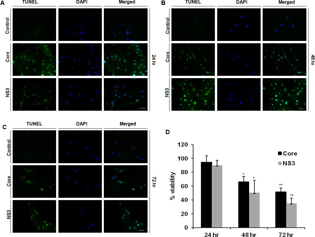
 Figure 6 of
Rajalakshmy, Mol Vis 2014; 20:1388-1397.
Figure 6 of
Rajalakshmy, Mol Vis 2014; 20:1388-1397.