Figure 9. Colocalization of RIP3 and eRAGE with Brn3a-labeled retinal ganglion cells in the IR-injured rat retina at the 12 h post-ischemic
reperfusion time point. A: The transcription factor BRN3a (green) is present in the ganglion cells of the retinal ganglion cell (RGC) layer. B: RIP3 (red) is localized to cells in the RGC layer in the ischemia reperfusion (IR)-injured rat retina. C: Double immunolabeling shows Brn3a (green) and RIP3 (red) are colocalized. D: eRAGE (red) is localized to cells in the RGC layer in the IR-injured retina. E: RIP3 (green) is localized to cells in the RGC layer in the IR-injured retina F: Double immunolabeling shows eRAGE (red) and RIP3 (green) are colocalized in the IR-injured retina. G: BRN3a (green) in the healthy rat retina (control). H: RIP3 (red) in the healthy rat retina (control). I: Double immunolabeling with Brn3a (green) and RIP3 (red) in the healthy rat retina (control). Scale bars=20 μm.
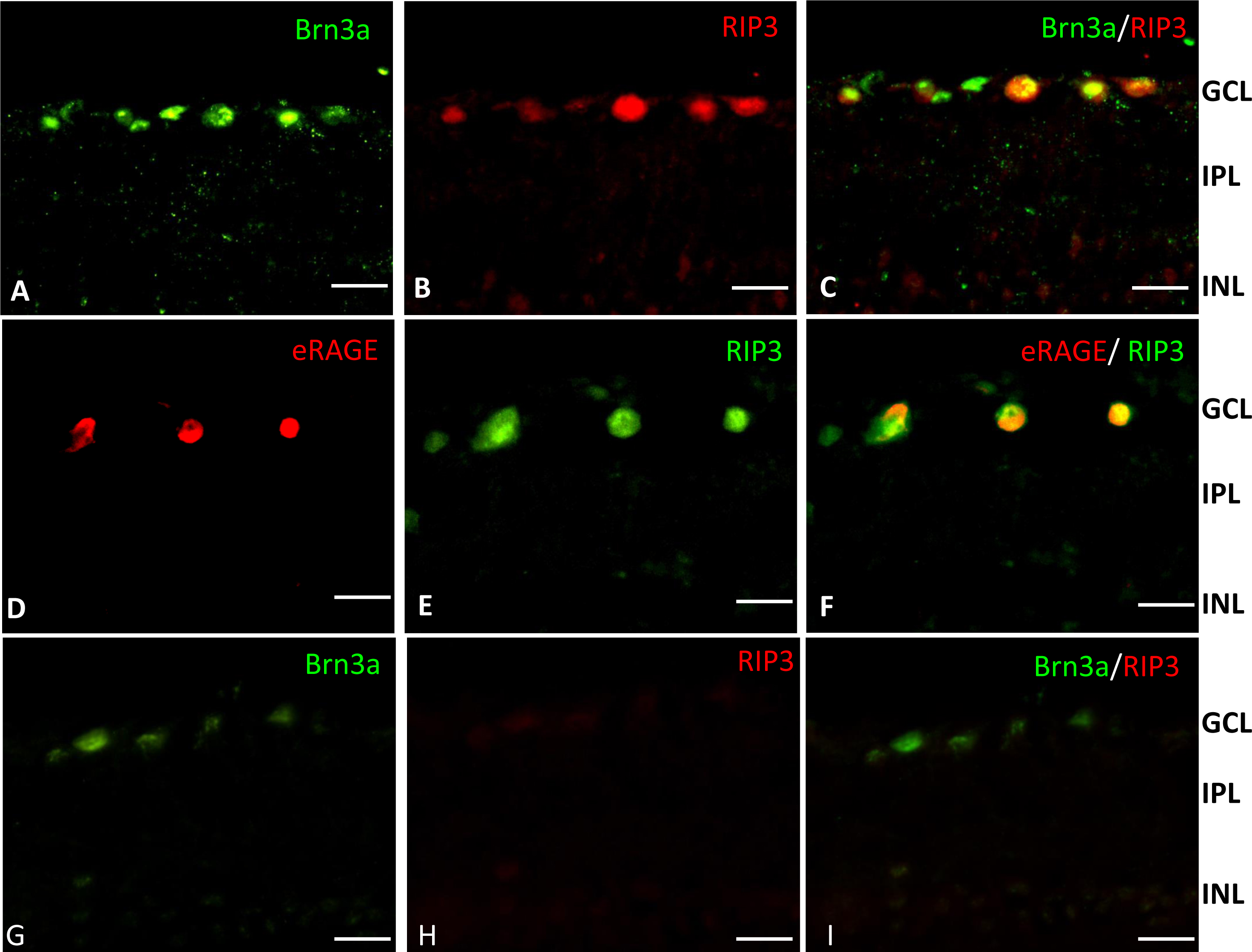
 Figure 9 of
Gao, Mol Vis 2014; 20:1374-1387.
Figure 9 of
Gao, Mol Vis 2014; 20:1374-1387.