Figure 7. Western blots indicating accumulation of p-ERK and t-ERK in the IR-injured rat retina. A: There was little difference in total ERK (t-ERK) abundance at each time point in the experimental and sham control groups.
However, the phosphorylated ERK protein (p-ERK) levels were increased at 12 h post-ischemia. The western blot is representative
of the blots for each of the three animals. B: At 12 h post-ischemia reperfusion, the p-ERK accumulation was significantly increased relative to the sham controls and
compared to all other time points. There was a gradual decrease in phosphorylated ERK with time after the 12 h ischemia reperfusion
(IR) injury period. Data are presented as mean ± standard error of the mean (SEM), *p<0.05, ***p<0.001. E=experimental group;
S=sham control group.
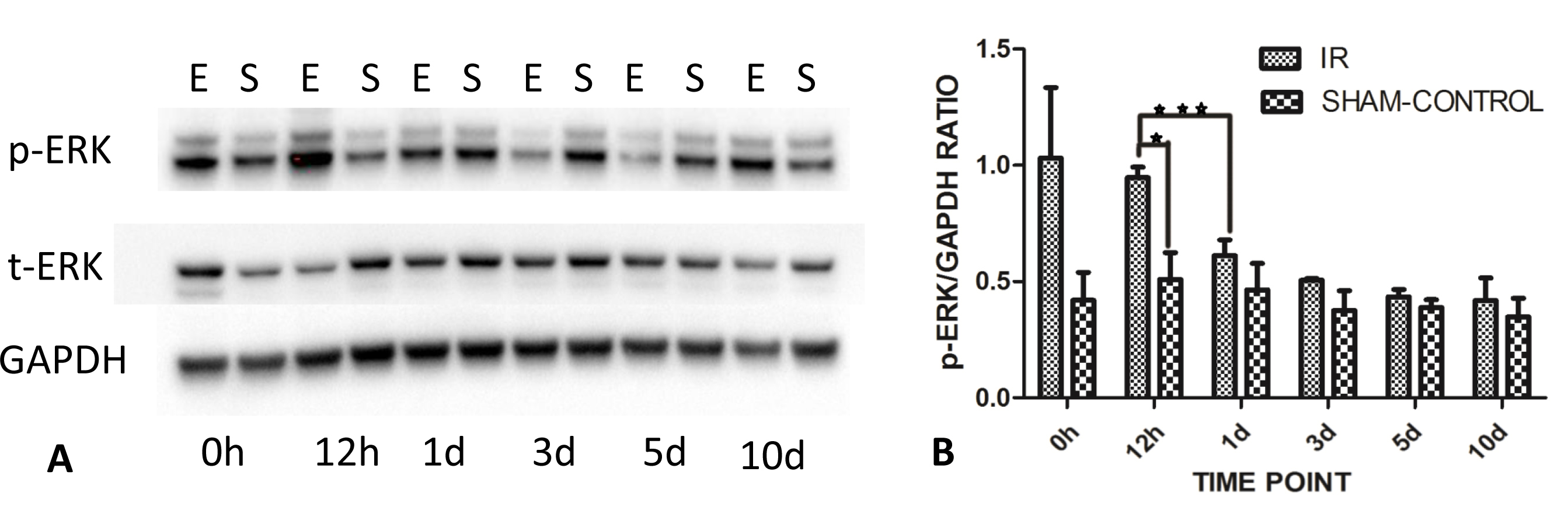
 Figure 7 of
Gao, Mol Vis 2014; 20:1374-1387.
Figure 7 of
Gao, Mol Vis 2014; 20:1374-1387.