Figure 6. Localization of the RAGE protein in the retina using the two anti-RAGE antibodies at 12 h post-ischemia. A: The Abcam-anti-RAGE antibody (green) localized the cRAGE protein in the nucleus only. B: The R&D-anti-RAGE antibody (red) localized the eRAGE protein in the cytoplasm and the nucleus. C: eRAGE and cRAGE colocalized in cells of the retinal ganglion layer (RGC) layer at the 12 h post-ischemic time point. The
colocalization shows the nucleus contains cRAGE and eRAGE. Scale bars=20 μm.
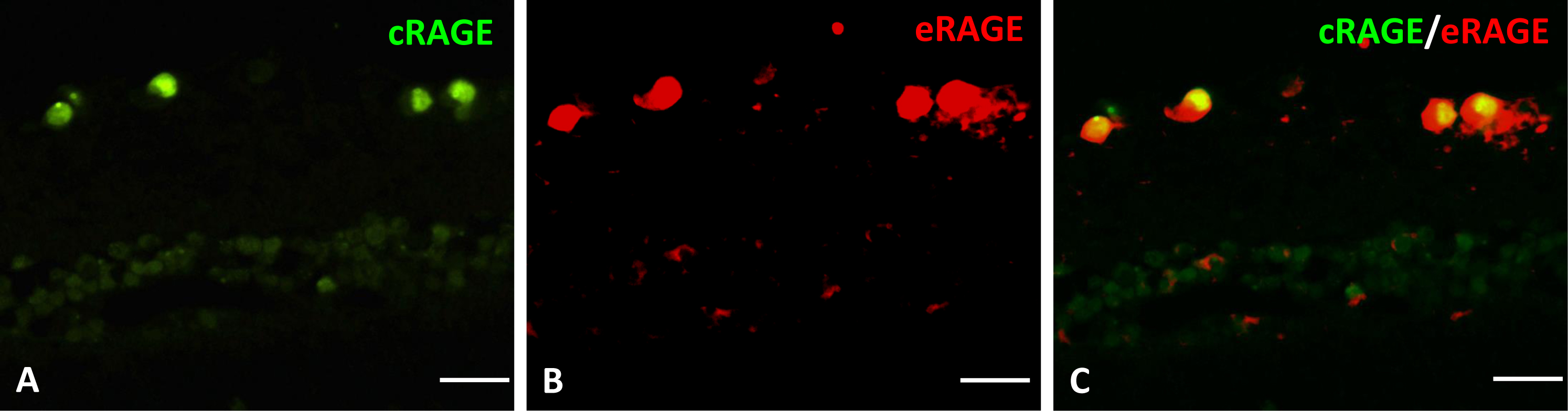
 Figure 6 of
Gao, Mol Vis 2014; 20:1374-1387.
Figure 6 of
Gao, Mol Vis 2014; 20:1374-1387.