Figure 3. Immunohistochemical staining of the RAGE protein in the rat retina. A: The eRAGE protein is absent in the naïve (wild-type, WT) rat retina. B: The eRAGE protein accumulates in the retinal ganglion layer (RGC) layer of the retina at the 12 h post-ischemic time point.
C: The eRAGE protein is not present in the retina at the 1 d post-ischemia-reperfusion period. The R&D anti-RAGE antibody (red)
was used. Scale bars=20 μm.
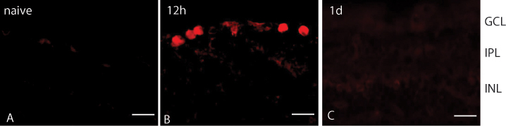
 Figure 3 of
Gao, Mol Vis 2014; 20:1374-1387.
Figure 3 of
Gao, Mol Vis 2014; 20:1374-1387.