Figure 2. Western blots indicating the presence of the RAGE protein in the retina of the IR-injured rat. Six time points are presented
on each blot. Every time point contained the experimental group (E) and the sham control group (S). A: The R&D-anti-RAGE antibody detected increased eRAGE accumulation at 12 h post ischemia. B: The peptide competition assay confirmed the specific band reactivity of the R&D-anti-RAGE antibody; lane-a western blot with R&D-anti-RAGE antibody preincubated with blocking peptide; lane-b western blot with a R&D-anti-RAGE antibody not preincubated with peptide. C: Western blots with R&D-anti-RAGE antibody performed under reducing and non-reducing conditions; lane-a non-reducing condition; lane-b reducing condition (β-mercaptoethanol); lane-c reducing condition (β-mercaptoethanol and dithiothreitol (DTT)). D: Abcam-anti RAGE-antibody shows the presence of cRAGE in every post-ischemic time point in the experimental and sham control
animals. No significant difference in the accumulation of the cRAGE protein was detected among the examined post-ischemic
time points.
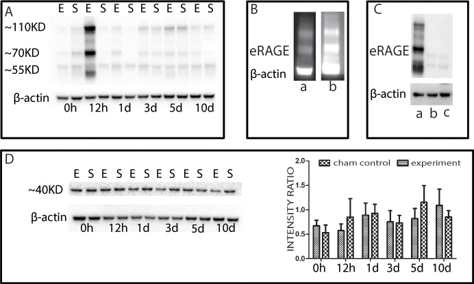
 Figure 2 of
Gao, Mol Vis 2014; 20:1374-1387.
Figure 2 of
Gao, Mol Vis 2014; 20:1374-1387.