Figure 10. Western blots indicating the presence of two FLIP isoforms, FLIPL and FLIPS, in the retina of the IR-injured rat. The FLIPS/L protein was strongly expressed only at the 12 h post-ischemic time point. Although weak FLIPL bands are visible, the FLIPS protein was detected only at the 12 h post- ischemic time point. E=experimental group; S=sham control group.
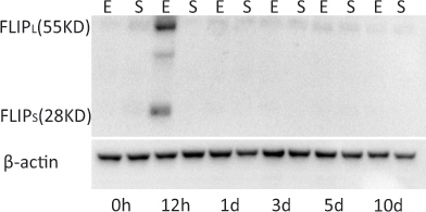
 Figure 10 of
Gao, Mol Vis 2014; 20:1374-1387.
Figure 10 of
Gao, Mol Vis 2014; 20:1374-1387.