Figure 9. nAChR IHC in α7 nAChR KO and WT mouse retinas. (A) Labeling patterns of the α3/α5 nAChR antibody (red) revealed labeling in the amacrine (arrows), bipolar (arrowheads), and
ganglion cells (asterisks) in both the WT and α7 nAChR KO mouse retinas, with less intense labeling in the KO mouse retinas.
Inner plexiform layer (IPL) immunoreactivity encompassed ChAT (green) immunoreactive IPL bands, but there were no double labeled
ChAT-positive cell bodies.
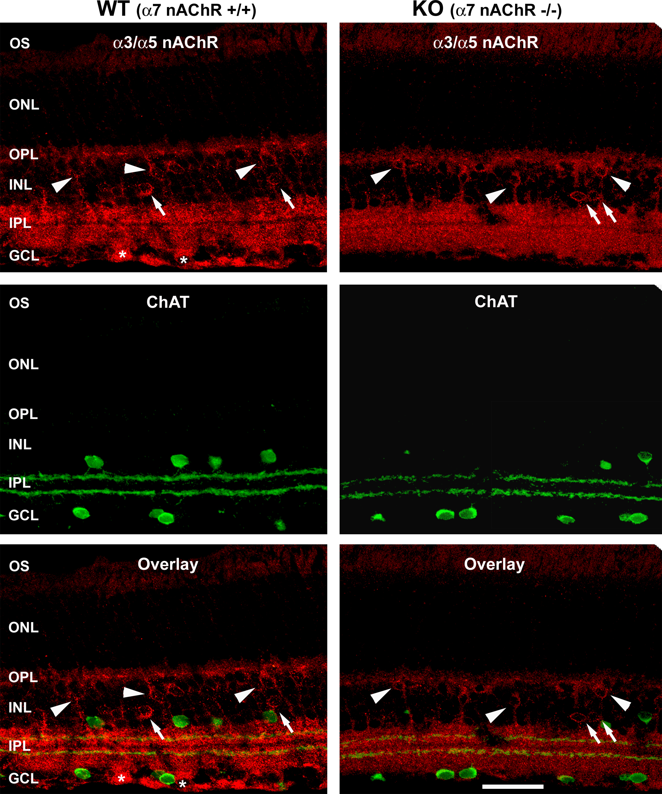
 Figure 9 of
Smith, Mol Vis 2014; 20:1328-1356.
Figure 9 of
Smith, Mol Vis 2014; 20:1328-1356.