Figure 4. Images of vertical sections depicting laser capture microdissection (LCM) of the unfixed mouse retinas. Vertical section of
an unfixed mouse retina. A: before LCM; B; after dissection of the ganglion cell layer (GCL–); C: after dissection of the GCL and the inner portion of the inner nuclear layer (iINL–), and D: after dissection of the GCL, the iINL, and the outer portion of the inner nuclear layer (oINL–). Scale bar, 100 μm.
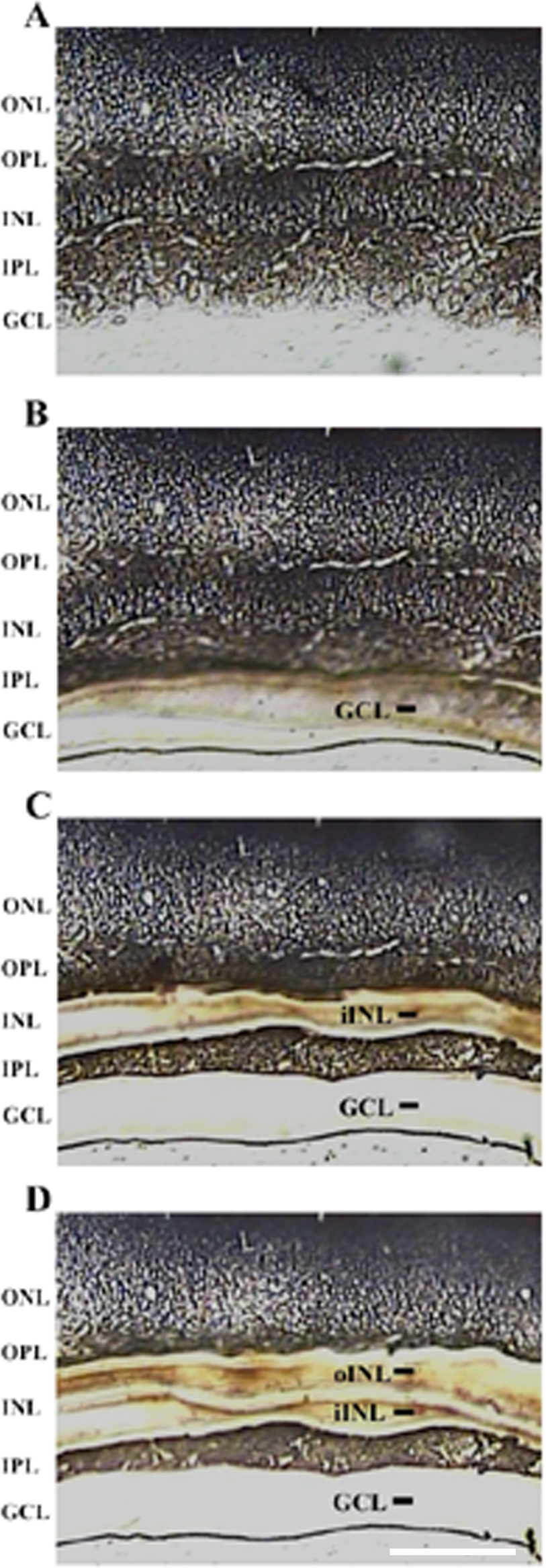
 Figure 4 of
Smith, Mol Vis 2014; 20:1328-1356.
Figure 4 of
Smith, Mol Vis 2014; 20:1328-1356.