Figure 11. mAChR ICH in α7 nAChR KO and WT mouse retinas. (A) Labeling patterns of the m1 mAChR antibody (red) revealed labeling in ganglion cells (asterisks) in the WT and the α7 nAChR
KO mouse retinas. There was broad diffuse IPL labeling in the IPL of the α7 nAChR KO mouse retinas as well as two bands of
immunoreactivity (double asterisks) that were not evident in the IPL of the WT mouse retinas. The α9 immunoreactive IPL bands
were directly beneath but did not colocalize with the ChAT (green) immunoreactive bands. (B) Labeling patterns of the m2 mAChR antibody (red) revealed labeling in the ganglion cells (asterisks) and labeling throughout
the IPL of the WT mouse retinas. IPL labeling was more intense in in sublaminae 2 and 3 and colocalized with ChAT immunoreactivity
in sublamina 2. There were no labeled cell bodies in the INL of the WT mouse retinas. In contrast, both bipolar (arrowheads)
and amacrine cells, including cholinergic amacrine cells (arrows), were immunoreactive for m2 in the α7 nAChR KO mouse retinas.
The brighter bands (double asterisks) within the IPL at sublaminae 2 and 3 were more intense in the α7 nAChR KO mouse retinas,
and the colocalization with the ChAT (green) immunoreactive bands in sublamina 2 was more pronounced.
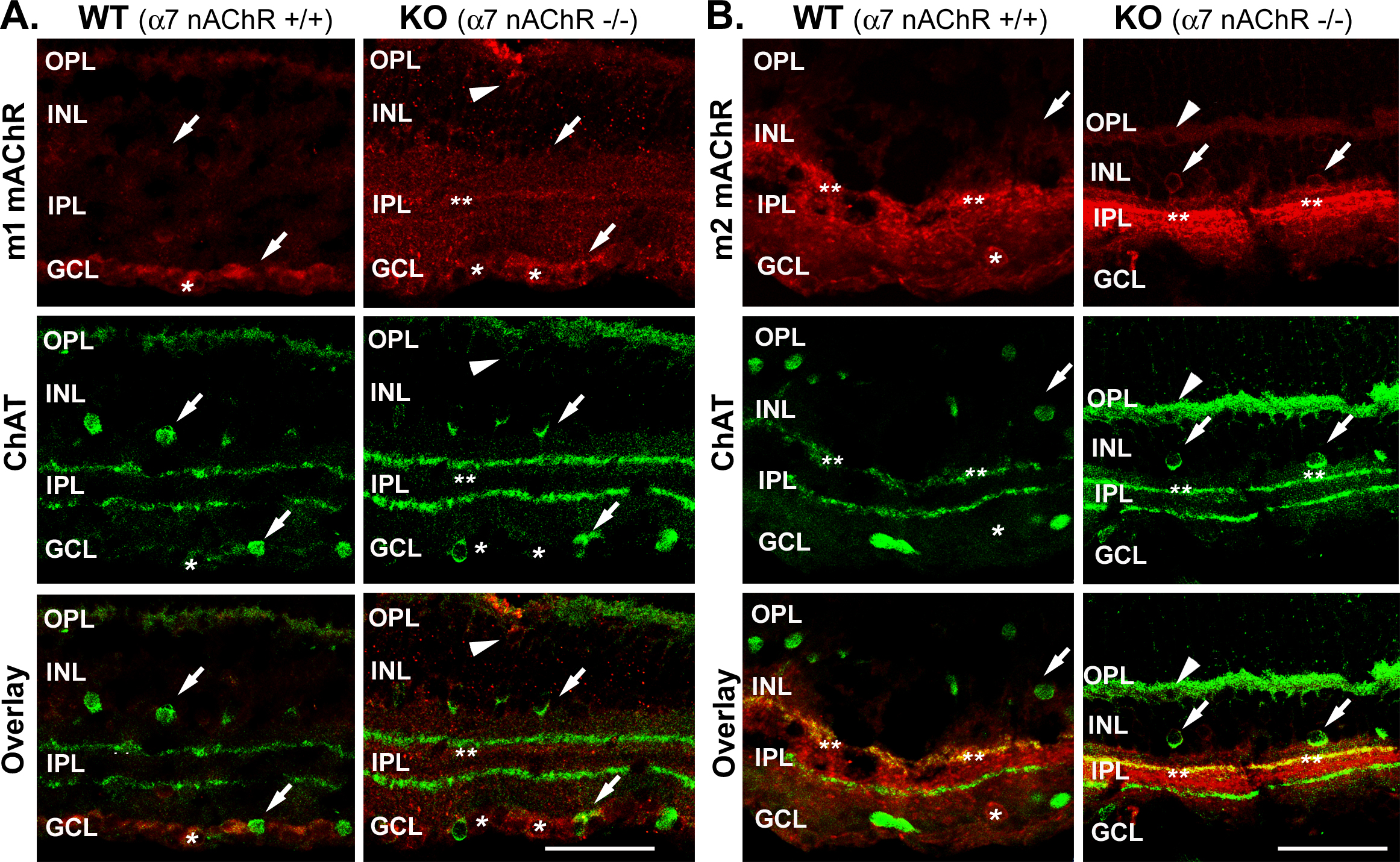
 Figure 11 of
Smith, Mol Vis 2014; 20:1328-1356.
Figure 11 of
Smith, Mol Vis 2014; 20:1328-1356.