Figure 10. Labeling patterns of the α9 nAChR antibody (red) revealed labeling in the amacrine (arrows), bipolar (arrowheads), and ganglion
cells (asterisks) with less cellular labeling in the α7 nAChR KO mouse retinas (right panels) than in the WT mouse retinas
(left panels). The α9 nAChR immunoreactivity was broadly distributed through the IPL and encompassed the ChAT (green) immunoreactive
bands. A subset of cholinergic amacrine cells demonstrated α9 immunoreactivity in the KO mouse retina. There were areas of
increased density at the margins and in the center of the IPL, particularly in the KO mouse retinas, at the same level as
the dim area in the center of the α3 and α5 nAChR immunoreactivity. Strong labeling in the outer plexiform layer (OPL) was
consistent with the labeling of bipolar cell dendrites.
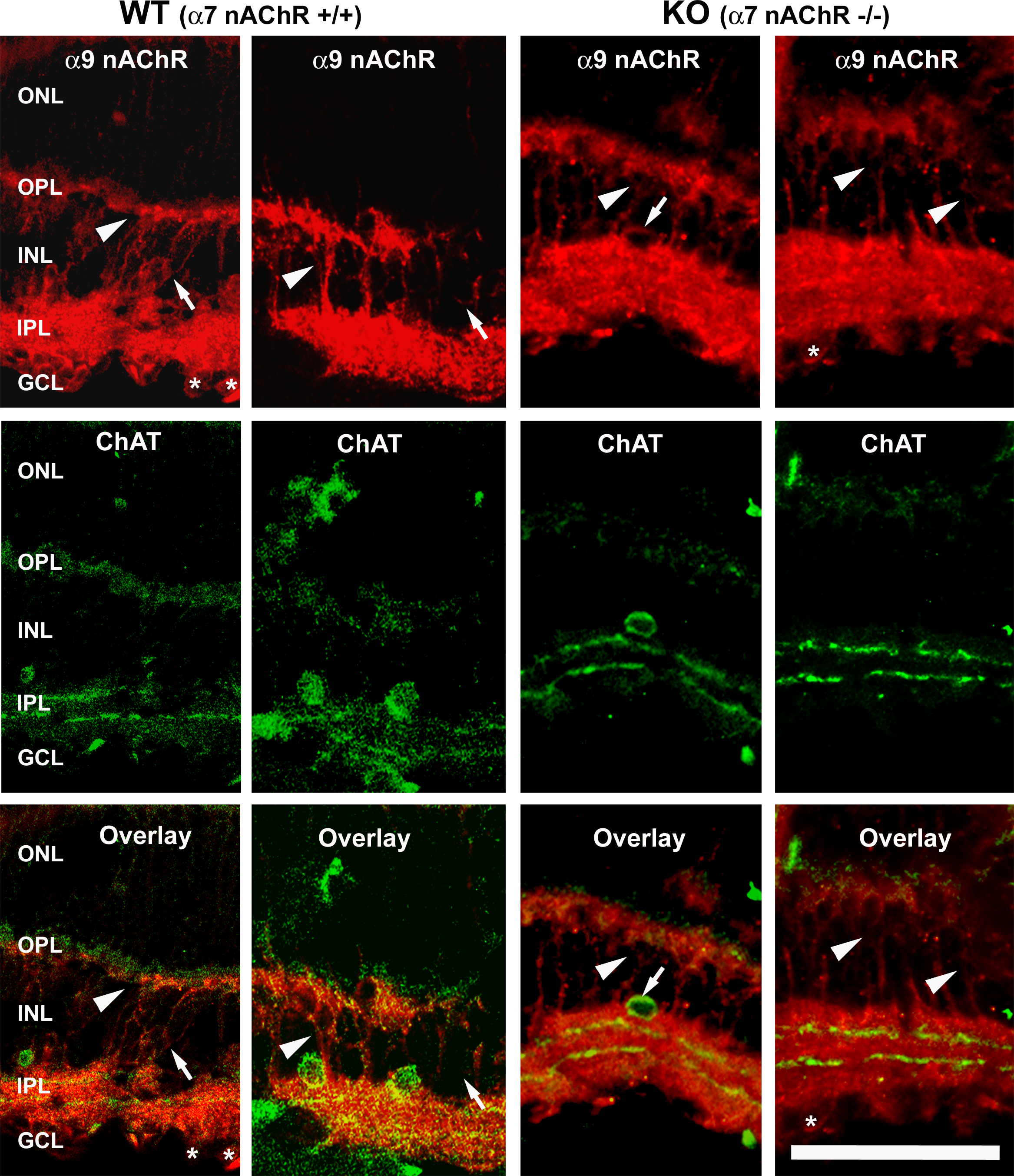
 Figure 10 of
Smith, Mol Vis 2014; 20:1328-1356.
Figure 10 of
Smith, Mol Vis 2014; 20:1328-1356.