Figure 3. Fundus photography and optical coherence tomography images of patient 113160. A: Fundus photography (top) and optical coherence tomography (OCT) image (bottom) of the right eye of patient 113160 shows
an unusually large retinoschisis cavity. B: His fundus photography (top) and OCT image (bottom) of the left eye.
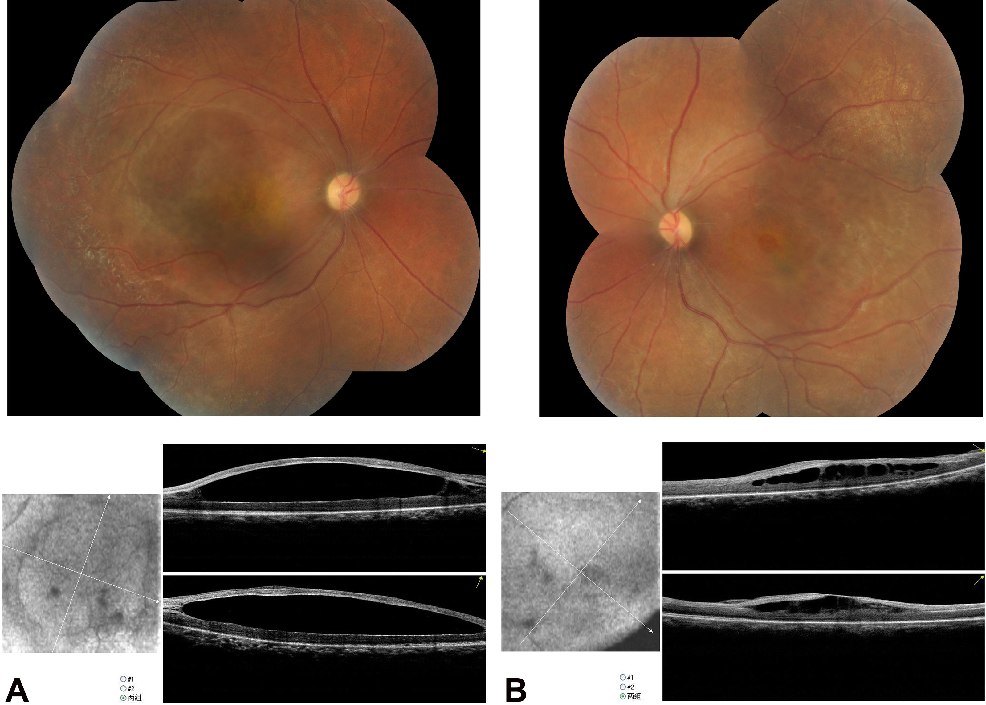
 Figure 3 of
Chen, Mol Vis 2014; 20:132-139.
Figure 3 of
Chen, Mol Vis 2014; 20:132-139.