Figure 2. Fundus photography and optical coherence tomography images of patients. A and B: Fundus appearance of patient 113001 shows foveal and peripheral retinal schisis in both eyes. C: Fundus photography of the left eye of patient 113010 shows a typical cystic-like foveal schisis. D: His optical coherence tomography (OCT) images show foveomacular schisis within the inner nuclear layer of the retina. E: Fundus photography of the left eye of patient 113100 shows normal foveal reflexes and a peripheral schisis cavity (arrow).
F: OCT images of patient 113100 show normal macular structure (top) and peripheral retinal schisis of the inner retina (bottom).
G: Fundus photography of the right eye of patient 110110 shows absent foveal reflexes. H: His OCT images present macular atrophy.
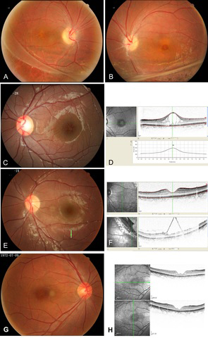
 Figure 2 of
Chen, Mol Vis 2014; 20:132-139.
Figure 2 of
Chen, Mol Vis 2014; 20:132-139.