Figure 3. Horizontal optical coherence tomography images cross sectioning the fovea in the right eyes of affected men and women who
carry a c.2543del mutation in RPGR ORF15. Choroidal thickness is indicated with a red bar. A: Normal retinal architecture in a 40-year-old control individual (enhanced depth imaging). B: Chorioretinal thinning, missing foveal depression, scar formation, and epiretinal membrane (arrow) in female III:1 at the
age of 64 years. C: Chorioretinal thinning, missing foveal depression, and vitreomacular traction associated with foveal elevation in female
III:4 at the age of 54 years. D: Chorioretinal thinning, myopic foveoschisis, and posterior staphyloma in female III:9 at the age of 50 years. E: Chorioretinal thinning and focal retinal thickening in male IV:1 at the age of 42 years. F: Chorioretinal thinning and myopic posterior staphyloma in male IV:2 at the age of 38 years. G: Chorioretinal thinning, macular hole, and myopic posterior staphyloma in male IV:3 at the age of 30 years. H: Normal retinal and choroidal architecture in female IV:4 at the age of 32 years (enhanced depth imaging). I: Choroidal thinning and normal retinal architecture in male IV:6 at the age of 19 years.
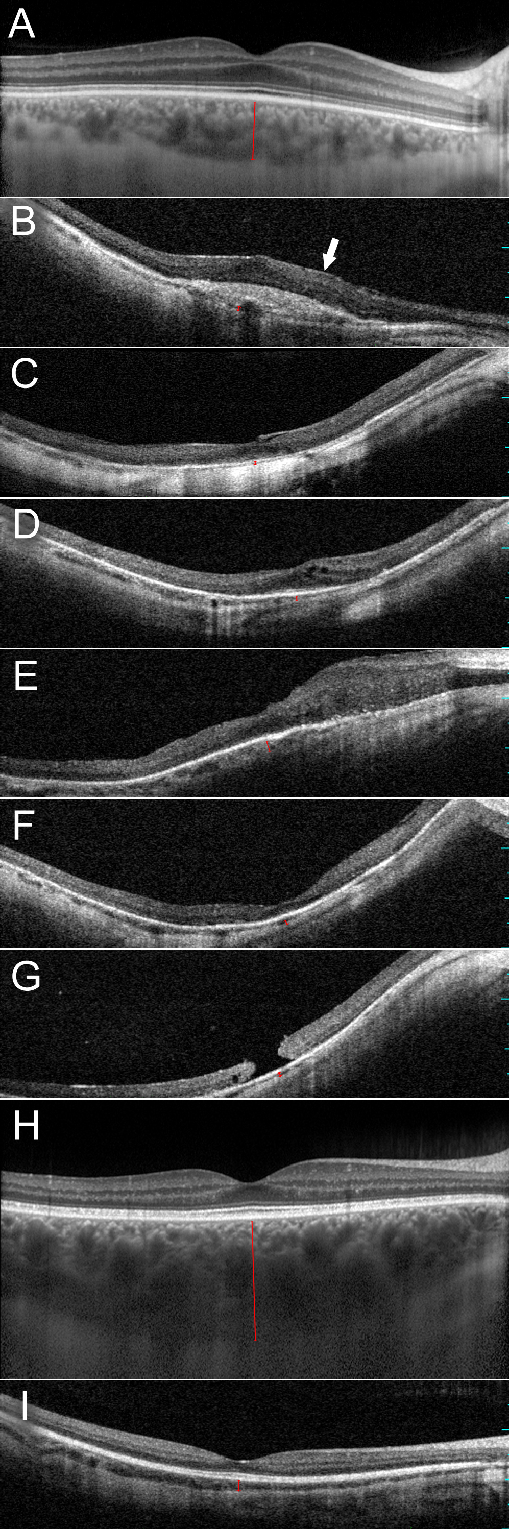
 Figure 3 of
Kousal, Mol Vis 2014; 20:1307-1317.
Figure 3 of
Kousal, Mol Vis 2014; 20:1307-1317.