Figure 8. Immunofluorescence staining of the TSPAN12 C189Y mutant. Cos 7 cells were transfected either with human wild-type or mutant TSPAN12 cloned into the pCMV6-entry vector,
or empty vector. Cells were washed with PBS after 48 h and fixed with 4% PFA for 15 min. Mouse monoclonal anti-Flag antibody
and Alexa Fluor 594 goat anti-mouse immunoglobulin (IgG) secondary antibody were used to detect TSPAN12 expression with the
standard immunostaining method. Red channel, TSPAN12; blue channel, 4',6-diamidino-2-phenylindole (DAPI) for nuclei staining.
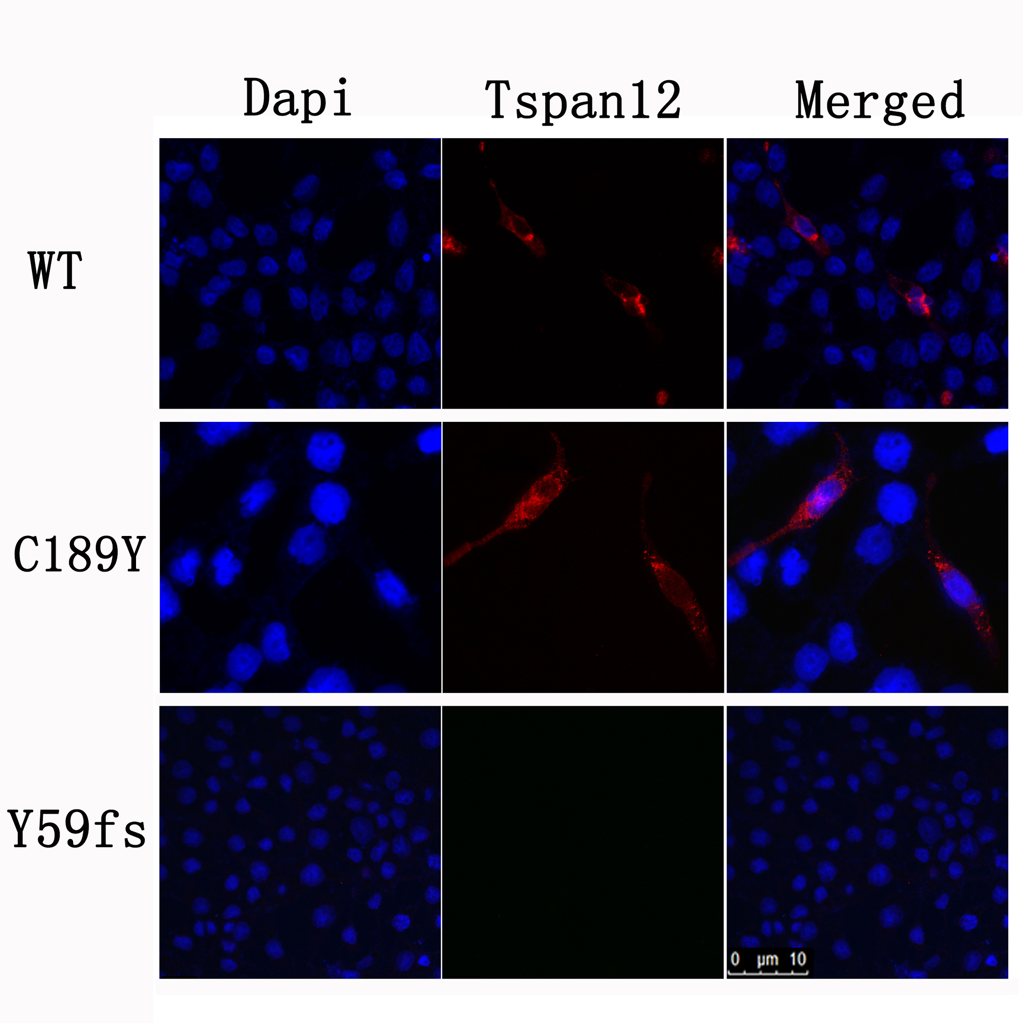
 Figure 8 of
Xu, Mol Vis 2014; 20:1296-1306.
Figure 8 of
Xu, Mol Vis 2014; 20:1296-1306.