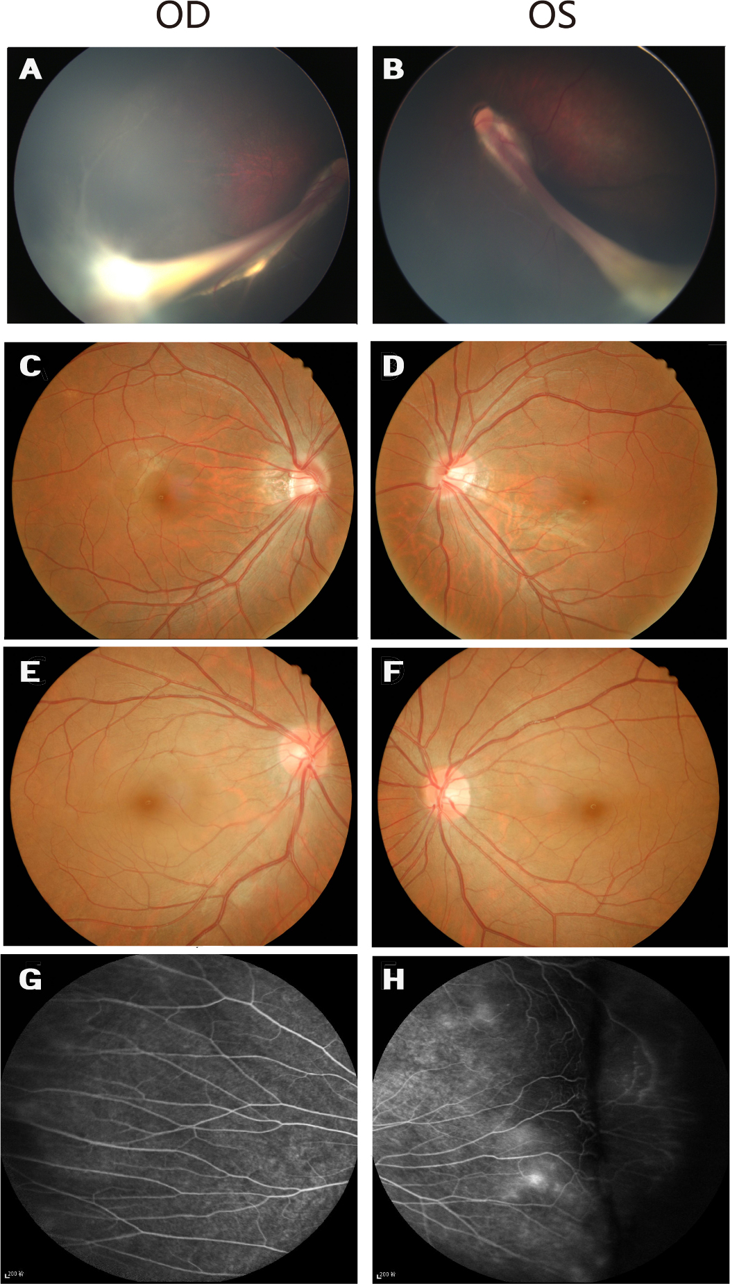
Figure 4. Fundus photographs of Family B with familial exudative vitreoretinopathy.
A and
B: Fundus photographs of the proband from Family B (individual II:1 in
Figure 1), showing the retinal vessels drawn up in a retinal fold that is obscuring the macula.
C and
D: The unaffected father has normal fundi.
E and
F: Fundus photographs of the asymptomatic mother with the c.177delC mutation show normal posterior fundi.
G and
H: The mother has increased vessel branching in the equatorial area and an avascular zone on the peripheral retina.
 Figure 4 of
Xu, Mol Vis 2014; 20:1296-1306.
Figure 4 of
Xu, Mol Vis 2014; 20:1296-1306.