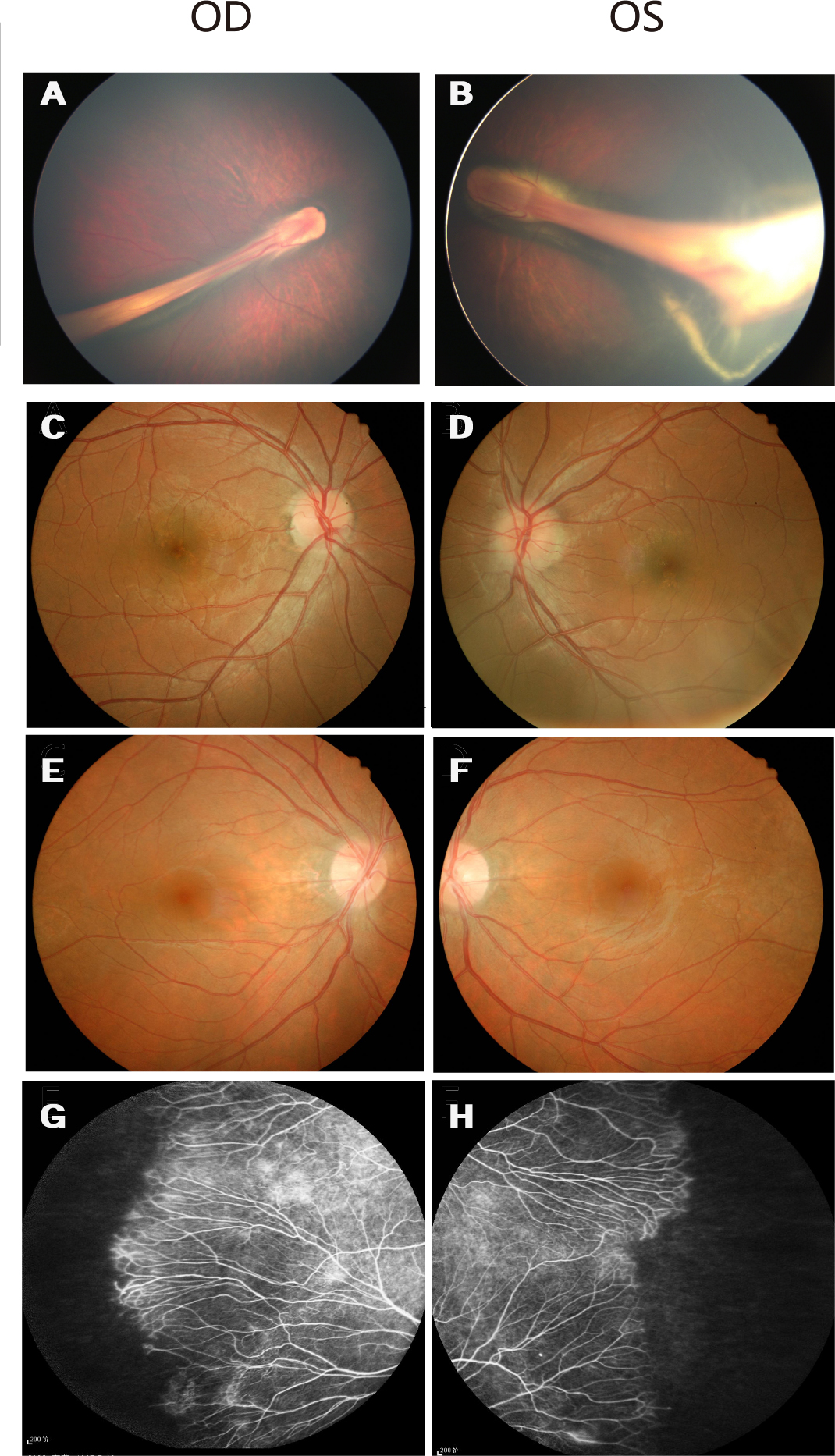
Figure 3. Fundus photographs of Family A with familial exudative vitreoretinopathy.
A and
B: Fundus photographs of the proband from Family A (individual II:1 in
Figure 1), showing a retinal fold and a dragged macula.
C and
D: The unaffected father without the mutation has normal fundi.
E and
F: Fundus photographs of the asymptomatic mother with the c.566G>A mutation show normal posterior fundi.
G and
H: The mother has areas of avascularity and abnormal vessels in the peripheral retina.
 Figure 3 of
Xu, Mol Vis 2014; 20:1296-1306.
Figure 3 of
Xu, Mol Vis 2014; 20:1296-1306.