Figure 6. Comparison of electroretinography recordings in the dark-adapted and light-adapted conditions. A: Dark-adapted (rod-mediated) electroretinography (ERG) responses. B: Light-adapted (cone-mediated) ERG responses. Electroretinography was performed on mice before the injections, 1 week, and
3 weeks after injections. The dark- and light-adapted ERG recordings were performed on the same mice. The ERG response amplitudes
in the Cyclosporin A (CsA) group were less reduced after the injection (p=0.114; p=0.01; in the dark-and-light-adapted conditions
respectively), when compared with the control group (p=0.040; p=0.04; in the dark-and-light-adapted conditions, respectively),
in both the dark-adapted and light-adapted conditions. However, the cone-mediated function in both groups was less affected
by the transplantation after 3 weeks than the rod-mediated function p = 0.008; p = 0.044, for the non-CsA and CsA, respectively).
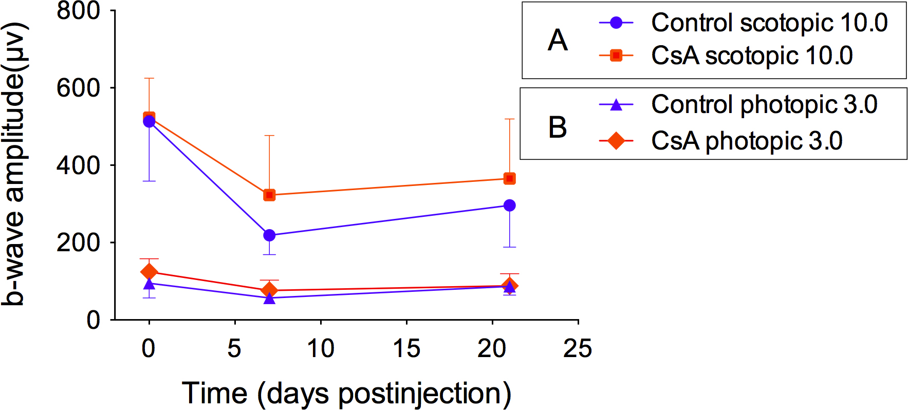
 Figure 6 of
Huang, Mol Vis 2014; 20:1271-1280.
Figure 6 of
Huang, Mol Vis 2014; 20:1271-1280.