Figure 7. Human retinal microvascular endothelial cells were left untreated (C) or treated with tumor necrosis factor-α (TNF-α) for
6 days. The expression levels of S100A4 and both intact and cleaved OPN were significantly increased in the TNF-α treated
samples compared to the controls. Western blot is representative of three different experiments, each performed in triplicate,
and bar graphs are representative of all three experiments. *The difference between the two means was statistically significant
at the 5% level.
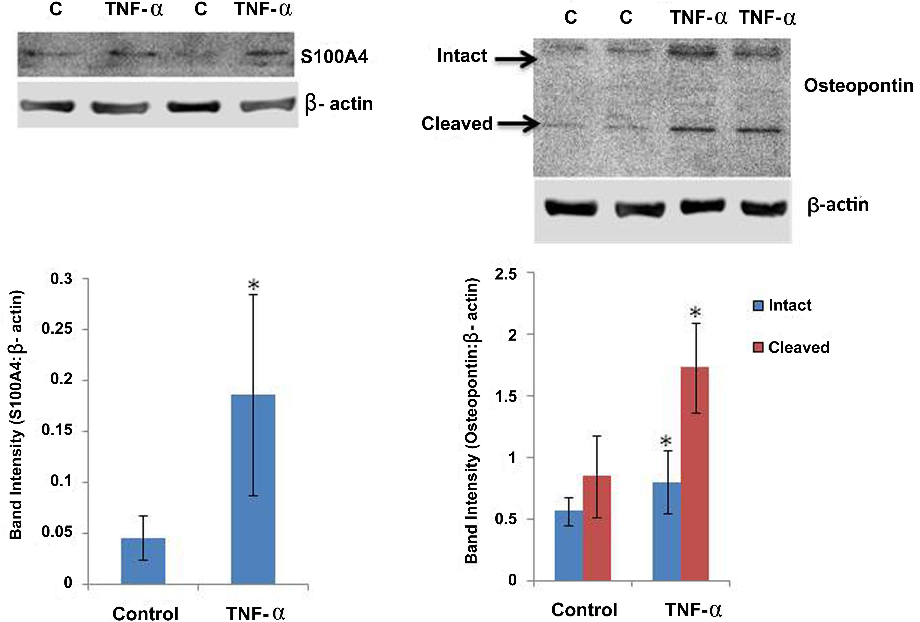
 Figure 7 of
Abu El-Asrar, Mol Vis 2014; 20:1209-1224.
Figure 7 of
Abu El-Asrar, Mol Vis 2014; 20:1209-1224.