Figure 6. Co-immunoprecipitation of the interaction between S100A4 and the receptor for advanced glycation end products (RAGE) in the
retina. Retinal tissue homogenates were immunoprecipitated using an antibody against S100A4 and normal rabbit IgG as a control
antibody. The relative abundance of RAGE in the S100A4 immunoprecipitates was determined by western blotting. The level of
S100A4 in the immunoprecipitated samples was used as an indicator of loading. Each experiment was repeated 3X with fresh samples
(n = 4). *The difference between the two means was statistically significant at the 5% level. IP = immunoprecipitation, IB
= immunoblotting, C = control, D = diabetic, IgG = normal rabbit IgG control antibody, Lysate = retinal tissue lysate.
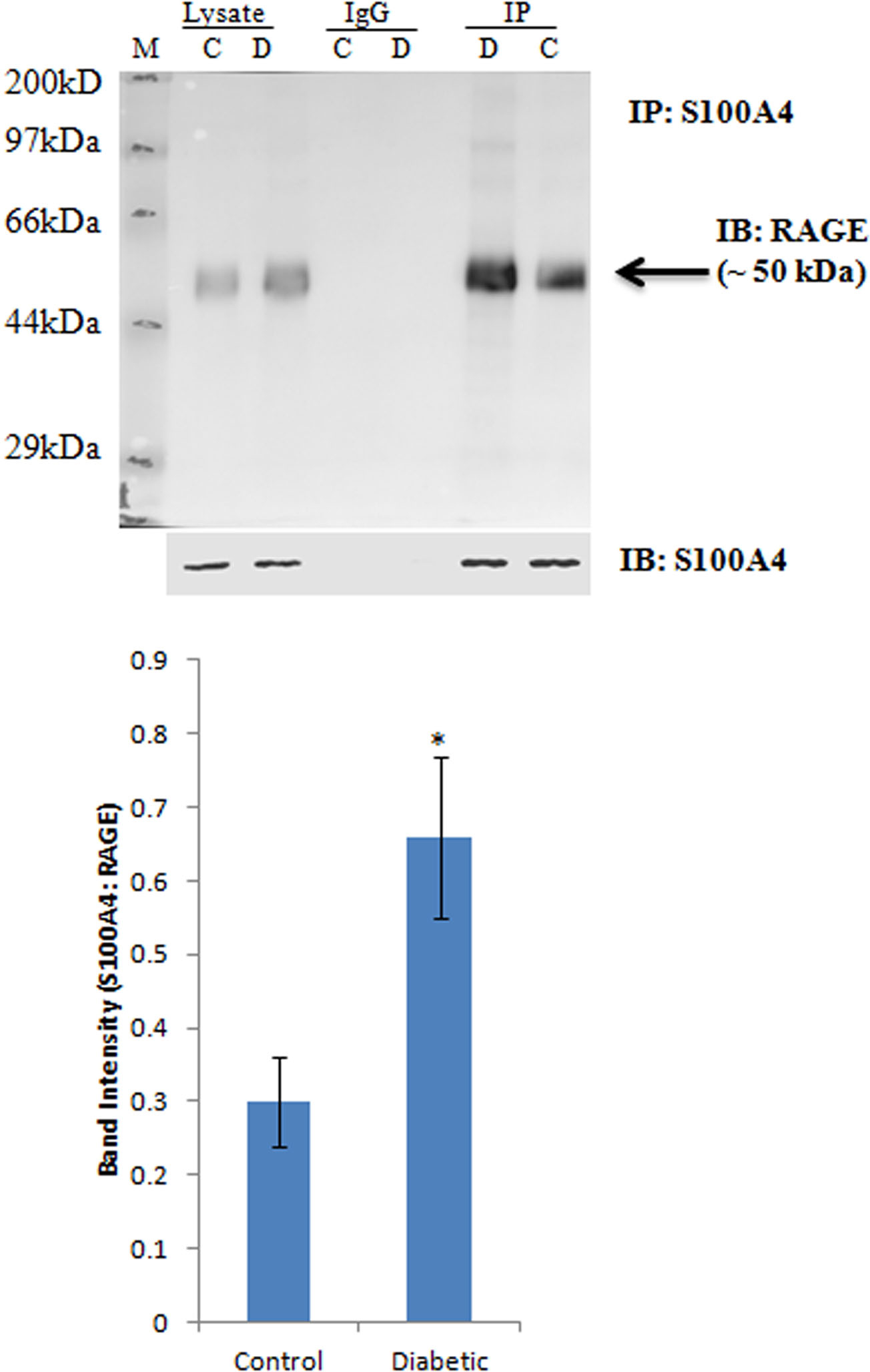
 Figure 6 of
Abu El-Asrar, Mol Vis 2014; 20:1209-1224.
Figure 6 of
Abu El-Asrar, Mol Vis 2014; 20:1209-1224.