Figure 4. PDR epiretinal membrane immunostainings. A negative control slide that was treated with an irrelevant antibody showing no
labeling (panel A; scale bar, 10 μm). Immunohistochemical staining for CD31 showing blood vessels positive for CD31 (panel B; scale bar, 10 μm). Immunohistochemical staining for S100A4 showing immunoreactivity in stromal cells (panel C; scale bar, 10 μm) and in the vascular endothelium (panel D; scale bar, 8 μm). Immunohistochemical staining for CD45 showing stromal cells positive for CD45 (panel E; scale bar, 10 μm). Double immunohistochemistry for CD45 (brown) and S100A4 (red) showing stromal cells co-expressing CD45
and S100A4 (arrows; panel F; scale bar, 10 μm).
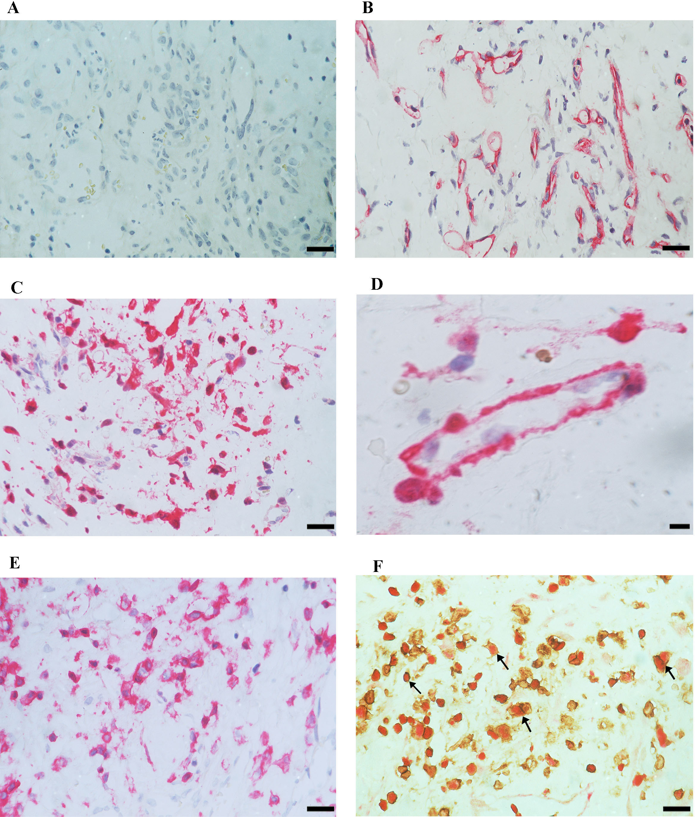
 Figure 4 of
Abu El-Asrar, Mol Vis 2014; 20:1209-1224.
Figure 4 of
Abu El-Asrar, Mol Vis 2014; 20:1209-1224.