Figure 1. Identification of differentially expressed proteins in pterygium compared to healthy conjunctiva. Crude extracts of healthy
conjunctiva as well as primary and recurrent pterygial tissues of eight subjects from each group were pooled; 50 μg of protein
from each pooled sample was separated with SDS–PAGE and stained with Coomassie brilliant blue. Fifteen bands of each sample
were excised and subjected to in-gel digestion. Digested peptides were then fractionated and identified with LC-MS/MS analysis.
Molecular weight markers are shown in the left side of the figure.
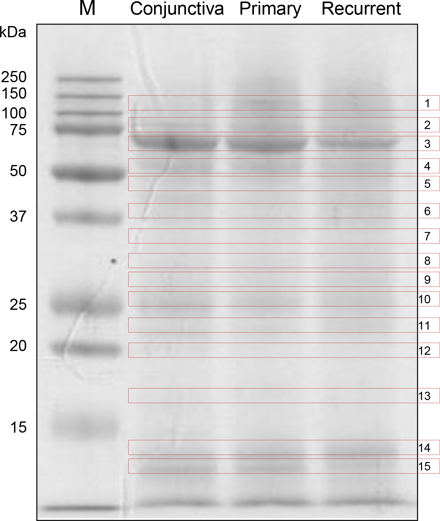
 Figure 1 of
Kim, Mol Vis 2014; 20:1192-1202.
Figure 1 of
Kim, Mol Vis 2014; 20:1192-1202.