Figure 7. Effect of chloroquine treatment on apoptosis during murine cytomegalovirus infection. Retinal pigment epithelial (RPE) cells
were infected with murine cytomegalovirus (MCMV) at low multiplicity of infection (MOI) = 1 in normal medium or in medium
containing chloroquine (10−6 M) for 2 and 3 days. A and B: Expression of cleaved caspase 3 was monitored and quantified. C: Collected cells were diluted to 1:1 using a 0.4% trypan blue solution. The stained cells and unstained cells were counted
under a microscope. The calculated percentage of unstained cells represents the percentage of viable cells. CQ: chloroquine;
2d: 2 days postinfection; 3d: 3 days postinfection. ***p<0.001, ANOVA. Data are shown as mean±SEM (n=3).
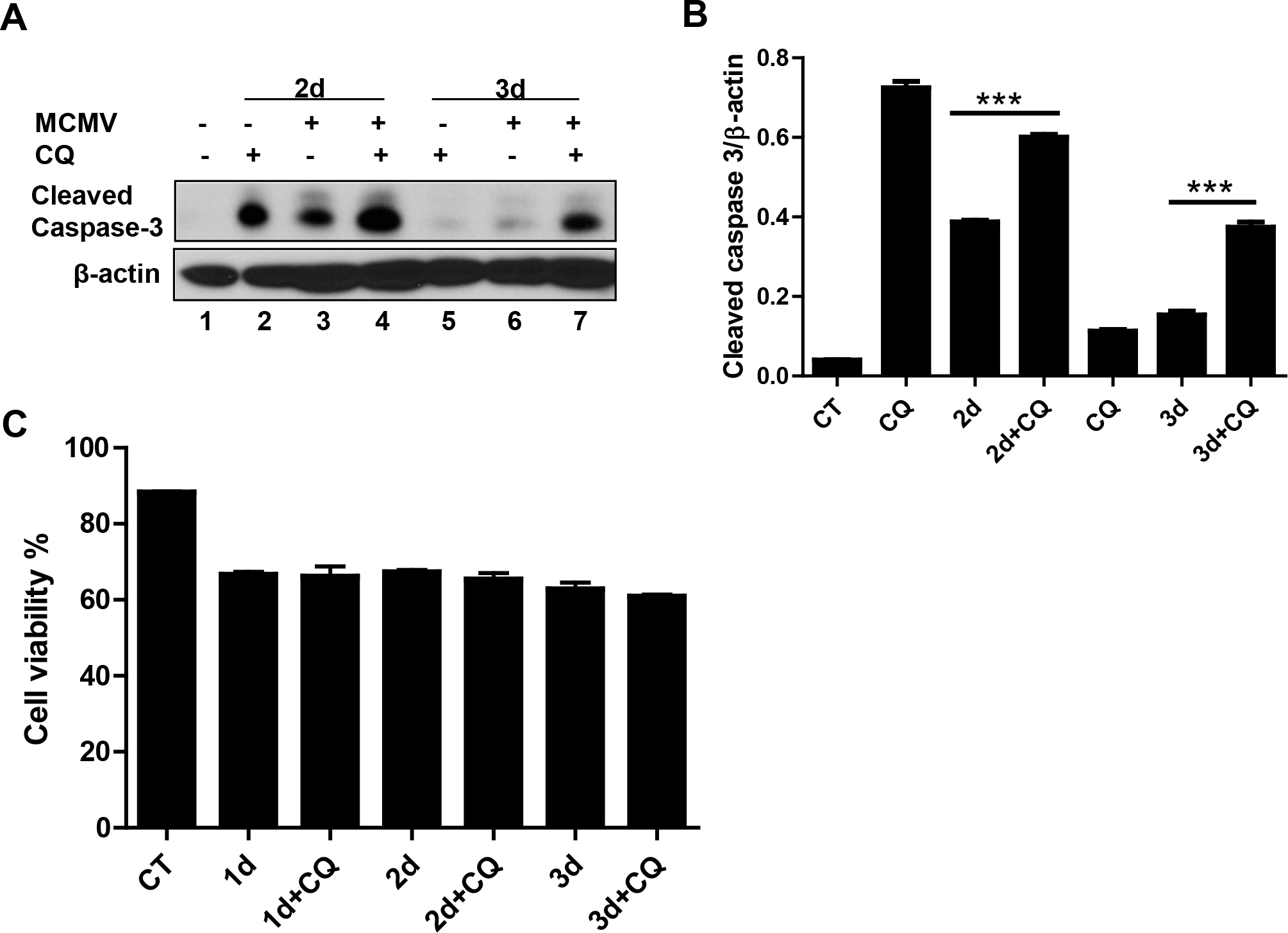
 Figure 7 of
Mo, Mol Vis 2014; 20:1161-1173.
Figure 7 of
Mo, Mol Vis 2014; 20:1161-1173.